- Phare
- Validé par KD/KO
Anticorps Polyclonal de lapin anti-CLOCK
CLOCK Polyclonal Antibody for WB, IHC, IF/ICC, ELISA
Hôte / Isotype
Lapin / IgG
Réactivité testée
Humain et plus (1)
Applications
WB, IHC, IF/ICC, ELISA
Conjugaison
Non conjugué
N° de cat : 18094-1-AP
Synonymes
Galerie de données de validation
Applications testées
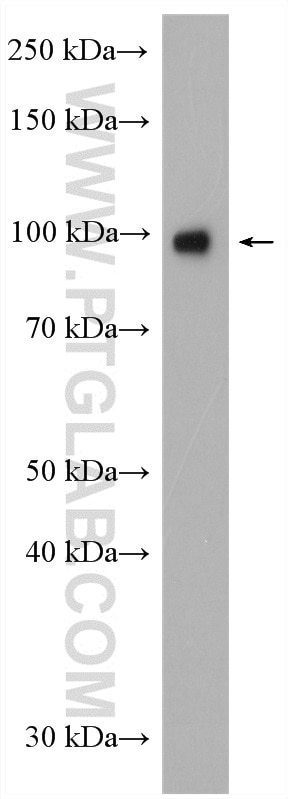
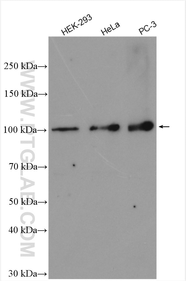
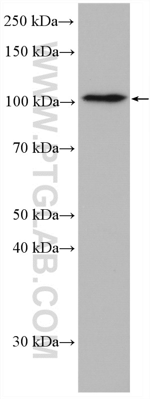
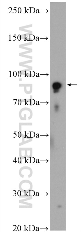
| Résultats positifs en WB | cellules HEK-293, cellules HeLa, cellules PC-3 |
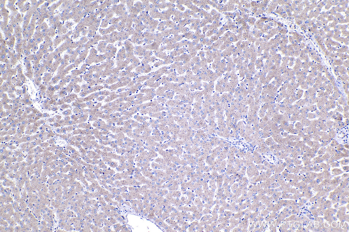
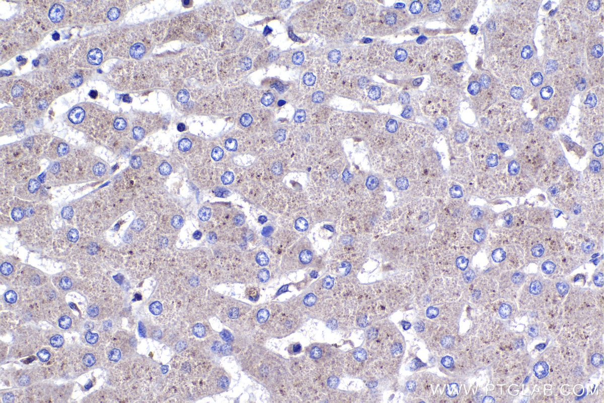
| Résultats positifs en IHC | tissu hépatique humain, il est suggéré de démasquer l'antigène avec un tampon de TE buffer pH 9.0; (*) À défaut, 'le démasquage de l'antigène peut être 'effectué avec un tampon citrate pH 6,0. |
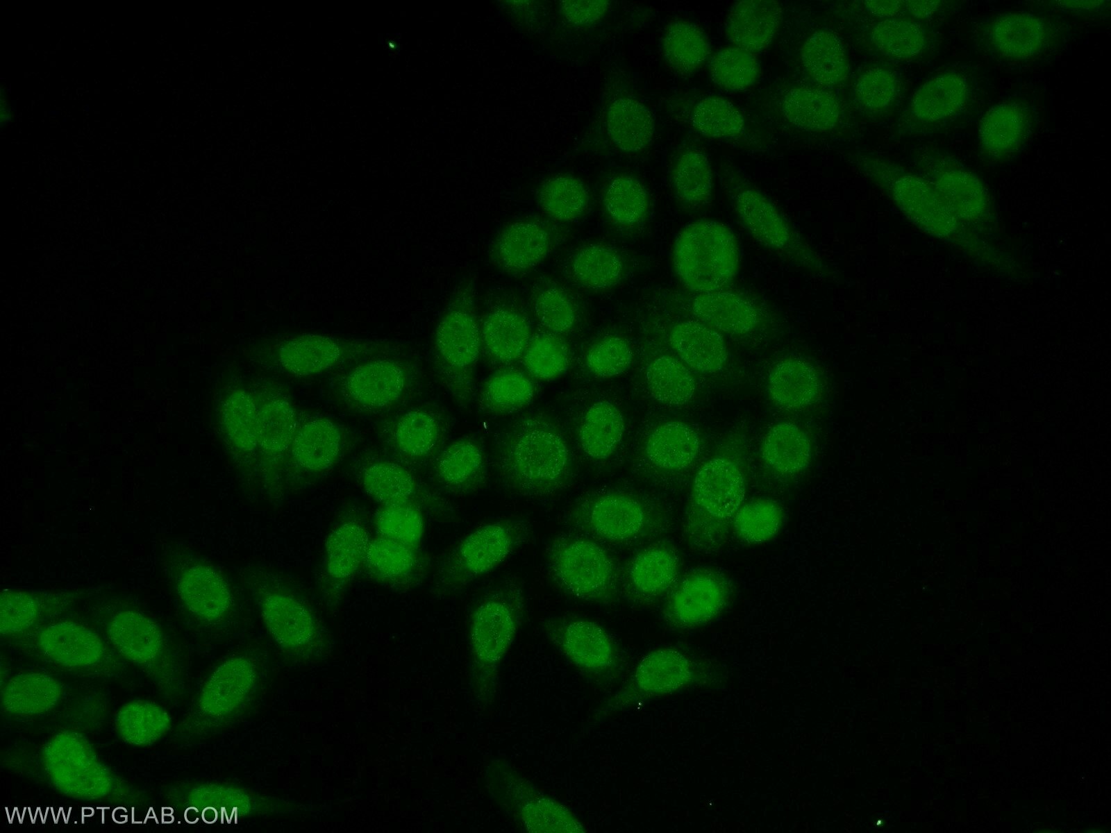
| Résultats positifs en IF/ICC | cellules HeLa |
Dilution recommandée
| Application | Dilution |
|---|---|
| Western Blot (WB) | WB : 1:500-1:2000 |
| Immunohistochimie (IHC) | IHC : 1:250-1:1000 |
| Immunofluorescence (IF)/ICC | IF/ICC : 1:20-1:200 |
| It is recommended that this reagent should be titrated in each testing system to obtain optimal results. | |
| Sample-dependent, check data in validation data gallery | |
Applications publiées
| KD/KO | See 1 publications below |
| WB | See 19 publications below |
| IHC | See 3 publications below |
| IF | See 1 publications below |
Informations sur le produit
18094-1-AP cible CLOCK dans les applications de WB, IHC, IF/ICC, ELISA et montre une réactivité avec des échantillons Humain
| Réactivité | Humain |
| Réactivité citée | Humain, souris |
| Hôte / Isotype | Lapin / IgG |
| Clonalité | Polyclonal |
| Type | Anticorps |
| Immunogène | CLOCK Protéine recombinante Ag12826 |
| Nom complet | clock homolog (mouse) |
| Masse moléculaire calculée | 846 aa, 95 kDa |
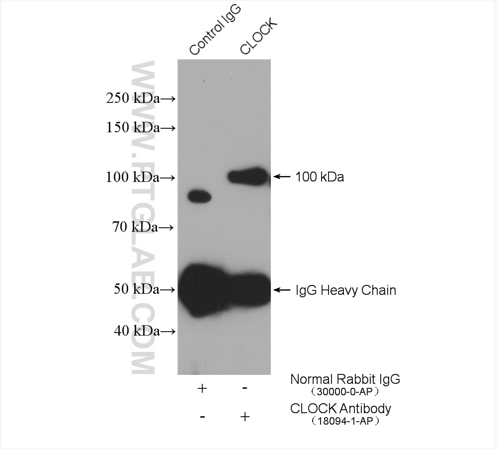
| Poids moléculaire observé | 95-110 kDa |
| Numéro d’acquisition GenBank | BC041878 |
| Symbole du gène | CLOCK |
| Identification du gène (NCBI) | 9575 |
| Conjugaison | Non conjugué |
| Forme | Liquide |
| Méthode de purification | Purification par affinité contre l'antigène |
| Tampon de stockage | PBS with 0.02% sodium azide and 50% glycerol |
| Conditions de stockage | Stocker à -20°C. Stable pendant un an après l'expédition. L'aliquotage n'est pas nécessaire pour le stockage à -20oC Les 20ul contiennent 0,1% de BSA. |
Informations générales
Circadian locomoter output cycles protein kaput (CLOCK), also named as BHLHE8 or KIAA0334, is a 846 amino acid protein, which contains one bHLH domain, one PAC domain, and two PAS domains. CLOCK localizes in the nucleus and cytoplasm. CLOCK) is expressed in all tissues examined including spleen, thymus, prostate, testis, ovary, small intestine, colon, leukocytes, heart, brain, placenta, lung, liver, skeletal muscle, kidney and pancreas. ARNTL/2-CLOCK heterodimers activate E-box element (5'-CACGTG-3') transcription of a number of proteins of the circadian clock, such as PER1 and PER2. The calculated molecular weight of CLOCK is 95kDa, we detected a 95-110 kDa protein by western blot. Our result is similar to the result of reseach paper (PMID: 30683868).
Protocole
| Product Specific Protocols | |
|---|---|
| WB protocol for CLOCK antibody 18094-1-AP | Download protocol |
| IHC protocol for CLOCK antibody 18094-1-AP | Download protocol |
| IF protocol for CLOCK antibody 18094-1-AP | Download protocol |
| IP protocol for CLOCK antibody 18094-1-AP | Download protocol |
| Standard Protocols | |
|---|---|
| Click here to view our Standard Protocols |
Publications
| Species | Application | Title |
|---|---|---|
Sci Total Environ Metastatic effects of perfluorooctanoic acid (PFOA) on Drosophila melanogaster with metabolic reprogramming and dysrhythmia in a multigenerational exposure scenario | ||
Int J Mol Sci Piperine Improves Lipid Dysregulation by Modulating Circadian Genes Bmal1 and Clock in HepG2 Cells.
| ||
J Agric Food Chem 3-MCPD Induced Mitochondrial Damage of Renal Cells Via the Rhythmic Protein BMAL1 Targeting SIRT3/SOD2 | ||
Neoplasia A long noncoding RNA perturbs the circadian rhythm of hepatoma cells to facilitate hepatocarcinogenesis. | ||
J Agric Food Chem Capsaicin Attenuates Oleic Acid-Induced Lipid Accumulation via the Regulation of Circadian Clock Genes in HepG2 Cells. |
Avis
The reviews below have been submitted by verified Proteintech customers who received an incentive for providing their feedback.
FH Verdiana (Verified Customer) (05-03-2022) | The bands are clear, there is no background noise even if the bands signal is not very strong. Good Ab.
|
