- Phare
- Validé par KD/KO
Anticorps Polyclonal de lapin anti-Cytochrome c
Cytochrome c Polyclonal Antibody for WB, IHC, IF-P, FC (Intra), ELISA
Hôte / Isotype
Lapin / IgG
Réactivité testée
Humain, rat, souris et plus (6)
Applications
WB, IHC, IF-P, FC (Intra), IP, ELISA
Conjugaison
Non conjugué
N° de cat : 10993-1-AP
Synonymes
Galerie de données de validation
 at dilution of 1:4000 incubated at room temperature for 1.5 hours.")
 with sh-Control and sh-Cytochrome c transfected HepG2 cells.")
 with sh-Control and sh-Cytochrome c transfected HEK-293 cells.")
 at dilution of 1:8000 incubated at room temperature for 1.5 hours.")
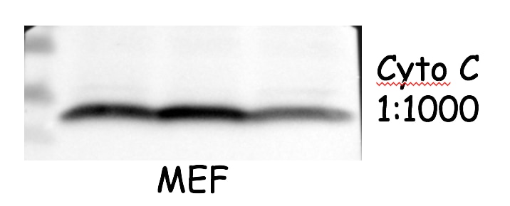
 at dilution of 1:1000 incubated at room temperature for 1.5 hours.")
 at dilution of 1:800 incubated at room temperature for 1.5 hours.")
 at dilution of 1:800 incubated at room temperature for 1.5 hours.")
 at dilution of 1:800 incubated at room temperature for 1.5 hours.")
 at dilution of 1:1000 (under 40x lens). Heat mediated antigen retrieval with Tris-EDTA buffer (pH 9.0).")
 at dilution of 1:1000 (under 10x lens). Heat mediated antigen retrieval with Tris-EDTA buffer (pH 9.0).")
 at dilution of 1:1000 (under 40x lens). Heat mediated antigen retrieval with Tris-EDTA buffer (pH 9.0).")
 at dilution of 1:1000 (under 10x lens). Heat mediated antigen retrieval with Tris-EDTA buffer (pH 9.0).")
 at dilution of 1:3000 (under 20x lens). Heat mediated antigen retrieval with Tris-EDTA buffer (pH 9.0).")
 at dilution of 1:1000 (under 10x lens). Heat mediated antigen retrieval with Tris-EDTA buffer (pH 9.0).")
 at dilution of 1:50 (under 10x lens).")
 at dilution of 1:50 (under 40x lens).")
 fixed paraffin-embedded mouse eye tissue using Cytochrome c antibody (10993-1-AP) at dilution of 1:200 and CoraLite®488-Conjugated AffiniPure Goat Anti-Rabbit IgG(H+L) (SA00013-2). Heat mediated antigen retrieval with Tris-EDTA buffer (pH 9.0).")
 fixed paraffin-embedded mouse eye tissue using Cytochrome c antibody (10993-1-AP) at dilution of 1:200 and CoraLite®488-Conjugated AffiniPure Goat Anti-Rabbit IgG(H+L) (SA00013-2). Heat mediated antigen retrieval with Tris-EDTA buffer (pH 9.0).")
 and CoraLite®488-Conjugated AffiniPure Goat Anti-Rabbit IgG(H+L) at dilution 1:1000 (red), or 0.4 ug Isotype Control. Cells were fixed with 4% PFA and permeabilized with Flow Cytometry Perm Buffer (PF00011-C).")
Applications testées
| Résultats positifs en WB | cellules HeLa, cellules C2C12, cellules HEK-293, cellules HepG2, cellules Jurkat, cellules NIH/3T3, tissu de muscle squelettique de rat, tissu de muscle squelettique de souris, tissu hépatique de rat, tissu rénal de rat, tissu rénal de souris |
| Résultats positifs en IHC | tissu hépatique humain, tissu cérébral de souris, tissu de cancer du côlon humain, tissu de cancer du sein humain il est suggéré de démasquer l'antigène avec un tampon de TE buffer pH 9.0; (*) À défaut, 'le démasquage de l'antigène peut être 'effectué avec un tampon citrate pH 6,0. |
| Résultats positifs en IF-P | tissu oculaire de souris, |
| Résultats positifs en FC (Intra) | cellules HepG2, |
Dilution recommandée
| Application | Dilution |
|---|---|
| Western Blot (WB) | WB : 1:1000-1:8000 |
| Immunohistochimie (IHC) | IHC : 1:500-1:2000 |
| Immunofluorescence (IF)-P | IF-P : 1:50-1:500 |
| Flow Cytometry (FC) (INTRA) | FC (INTRA) : 0.40 ug per 10^6 cells in a 100 µl suspension |
| It is recommended that this reagent should be titrated in each testing system to obtain optimal results. | |
| Sample-dependent, check data in validation data gallery | |
Applications publiées
| WB | See 582 publications below |
| IHC | See 33 publications below |
| IF | See 38 publications below |
| IP | See 1 publications below |
| FC | See 1 publications below |
Informations sur le produit
10993-1-AP cible Cytochrome c dans les applications de WB, IHC, IF-P, FC (Intra), IP, ELISA et montre une réactivité avec des échantillons Humain, rat, souris
| Réactivité | Humain, rat, souris |
| Réactivité citée | rat, Chèvre, Humain, porc, poulet, singe, souris, Hamster, Hippospongia |
| Hôte / Isotype | Lapin / IgG |
| Clonalité | Polyclonal |
| Type | Anticorps |
| Immunogène | Cytochrome c Protéine recombinante Ag1455 |
| Nom complet | cytochrome c, somatic |
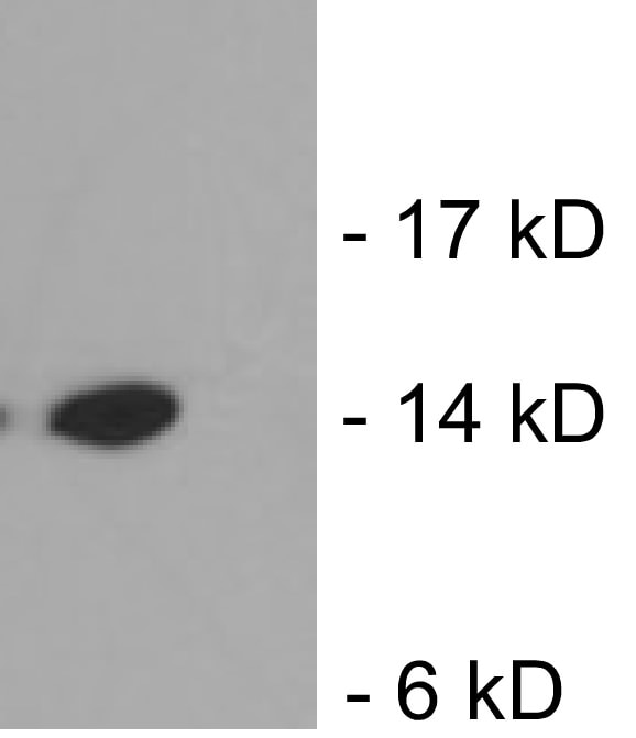
| Masse moléculaire calculée | 12 kDa |
| Poids moléculaire observé | 12-15 kDa |
| Numéro d’acquisition GenBank | BC009578 |
| Symbole du gène | Cytochrome c |
| Identification du gène (NCBI) | 54205 |
| Conjugaison | Non conjugué |
| Forme | Liquide |
| Méthode de purification | Purification par affinité contre l'antigène |
| Tampon de stockage | PBS with 0.02% sodium azide and 50% glycerol |
| Conditions de stockage | Stocker à -20°C. Stable pendant un an après l'expédition. L'aliquotage n'est pas nécessaire pour le stockage à -20oC Les 20ul contiennent 0,1% de BSA. |
Informations générales
Cytochrome c is a 12-15 kDa electron transporting protein located in the inner mitochondrial membrane. As a part of respiratory chain, cytochrome c plays a critical role in the process of oxidative phosphorylation and ATP producing. Besides, cytochrome c also gets implicated in apoptosis process. Upon apoptotic stimulation, cytochrome c ca99n be released from mitochondria into cytoplasm, which is required for caspase-3 activation and the occurrence of apoptosis.
Protocole
| Product Specific Protocols | |
|---|---|
| WB protocol for Cytochrome c antibody 10993-1-AP | Download protocol |
| IHC protocol for Cytochrome c antibody 10993-1-AP | Download protocol |
| IF protocol for Cytochrome c antibody 10993-1-AP | Download protocol |
| Standard Protocols | |
|---|---|
| Click here to view our Standard Protocols |
Publications
| Species | Application | Title |
|---|---|---|
Cell Tau interactome maps synaptic and mitochondrial processes associated with neurodegeneration. | ||
Nat Commun MYG1 drives glycolysis and colorectal cancer development through nuclear-mitochondrial collaboration | ||
Mol Cell Filamentous GLS1 promotes ROS-induced apoptosis upon glutamine deprivation via insufficient asparagine synthesis. | ||
Adv Sci (Weinh) Mitochondrial tRNAGlu 14693A>G Mutation, an "Enhancer" to the Phenotypic Expression of Leber's Hereditary Optic Neuropathy | ||
Adv Sci (Weinh) Hierarchical Targeting Nanodrug with Holistic DNA Protection for Effective Treatment of Acute Kidney Injury | ||
Acta Pharm Sin B Honokiol alleviated neurodegeneration by reducing oxidative stress and improving mitochondrial function in mutant SOD1 cellular and mouse models of amyotrophic lateral sclerosis |
Avis
The reviews below have been submitted by verified Proteintech customers who received an incentive for providing their feedback.
FH Kazu (Verified Customer) (12-14-2022) | Worked well for 4% PFA fixed mouse optic nerve using this antibody at dilution of 1:200. Little background observed.
|
FH Azita (Verified Customer) (06-02-2021) | Western blot analysis using Cytochrome c polyclonal antibody in NSC34 cell line at dilution of 1:1000.
|
FH Ying (Verified Customer) (04-22-2021) | good antibody. work well with mouse cells
 |
FH Chi (Verified Customer) (09-26-2019) | The antibody works very well with good signal and low background.
 |
FH Xiaoping (Verified Customer) (10-22-2018) | The signal is good and the size is between 10-15kDa.
 |

