Anticorps Polyclonal de lapin anti-p21
p21 Polyclonal Antibody for WB, IF/ICC, IP, ELISA
Hôte / Isotype
Lapin / IgG
Réactivité testée
rat, souris
Applications
WB, IHC, IF/ICC, IP, CoIP, ELISA
Conjugaison
Non conjugué
N° de cat : 28248-1-AP
Synonymes
Galerie de données de validation
 at dilution of 1:1000 incubated at room temperature for 1.5 hours.")
 at dilution of 1:800 incubated at room temperature for 1.5 hours.")
 with NIH/3T3 cells lysate 1920 ug.")
 fixed NIH/3T3 cells using 28248-1-AP (p21 antibody), at dilution of 1:200 and CoraLite®488-Conjugated AffiniPure Goat Anti-Rabbit IgG(H+L). F-actin is stained using CL555-phalloidin (red).")
 fixed NIH/3T3 cells using p21 antibody (28248-1-AP) at dilution of 1:200 and CoraLite®594-Conjugated Goat Anti-Rabbit IgG(H+L) (SA00013-4), CL488-phalloidin (green).")
Applications testées
| Résultats positifs en WB | cellules PC-12, cellules DC2.4, cellules HSC-T6, cellules Neuro-2a, cellules NIH/3T3, cellules RAW 264.7, cellules Sp2/0 |
| Résultats positifs en IP | cellules NIH/3T3, |
| Résultats positifs en IF/ICC | cellules NIH/3T3, |
Dilution recommandée
| Application | Dilution |
|---|---|
| Western Blot (WB) | WB : 1:500-1:2000 |
| Immunoprécipitation (IP) | IP : 0.5-4.0 ug for 1.0-3.0 mg of total protein lysate |
| Immunofluorescence (IF)/ICC | IF/ICC : 1:50-1:500 |
| It is recommended that this reagent should be titrated in each testing system to obtain optimal results. | |
| Sample-dependent, check data in validation data gallery | |
Applications publiées
| WB | See 76 publications below |
| IHC | See 16 publications below |
| IF | See 20 publications below |
| IP | See 1 publications below |
| CoIP | See 1 publications below |
Informations sur le produit
28248-1-AP cible p21 dans les applications de WB, IHC, IF/ICC, IP, CoIP, ELISA et montre une réactivité avec des échantillons rat, souris
| Réactivité | rat, souris |
| Réactivité citée | rat, souris |
| Hôte / Isotype | Lapin / IgG |
| Clonalité | Polyclonal |
| Type | Anticorps |
| Immunogène | p21 Protéine recombinante Ag28394 |
| Nom complet | cyclin-dependent kinase inhibitor 1A (P21) |
| Masse moléculaire calculée | 18 kDa |
| Poids moléculaire observé | 18 kDa |
| Numéro d’acquisition GenBank | NM_001111099 |
| Symbole du gène | p21 |
| Identification du gène (NCBI) | 12575 |
| Conjugaison | Non conjugué |
| Forme | Liquide |
| Méthode de purification | Purification par affinité contre l'antigène |
| Tampon de stockage | PBS with 0.02% sodium azide and 50% glycerol |
| Conditions de stockage | Stocker à -20°C. Stable pendant un an après l'expédition. L'aliquotage n'est pas nécessaire pour le stockage à -20oC Les 20ul contiennent 0,1% de BSA. |
Informations générales
CDKN1A (p21, CIP1, WAF1) is a cyclin-dependent kinase inhibitor. CDKN1A binds to and inhibits the activity of cyclin-CDK2 or -CDK4 complexes, and thus functions as a regulator of cell cycle progression at the G1 phase. The expression of CDKN1A is induced by wild-type but not mutant p53 protein, through which CDKN1A mediates the p53-dependent cell cycle G1 phase arrest in response to a variety of stress stimuli. CDKN1A can interact with proliferating cell nuclear antigen (PCNA), and plays a regulatory role in S phase DNA replication and DNA damage repair. CDKN1A was reported to be specifically cleaved by CASP3-like caspases, which thus leads to a dramatic activation of CDK2, and may be instrumental in the execution of apoptosis following caspase activation.
Protocole
| Product Specific Protocols | |
|---|---|
| WB protocol for p21 antibody 28248-1-AP | Download protocol |
| IF protocol for p21 antibody 28248-1-AP | Download protocol |
| IP protocol for p21 antibody 28248-1-AP | Download protocol |
| Standard Protocols | |
|---|---|
| Click here to view our Standard Protocols |
Publications
| Species | Application | Title |
|---|---|---|
Bioact Mater Chemo-immunotherapy by dual-enzyme responsive peptide self-assembling abolish melanoma | ||
Redox Biol Deficiency of S100 calcium binding protein A9 attenuates vascular dysfunction in aged mice | ||
EMBO J Kinesin-like motor protein KIF23 maintains neural stem and progenitor cell pools in the developing cortex | ||
Dev Cell Endoderm development requires centrioles to restrain p53-mediated apoptosis in the absence of ERK activity. | ||
Aging Cell Excessive processing and acetylation of OPA1 aggravate age-related hearing loss via the dysregulation of mitochondrial dynamics | ||
Biomed Pharmacother Seno-antigen-pulsed dendritic cell vaccine induce anti-aging immunity to improve adipose tissue senescence and metabolic abnormalities |
Avis
The reviews below have been submitted by verified Proteintech customers who received an incentive for providing their feedback.
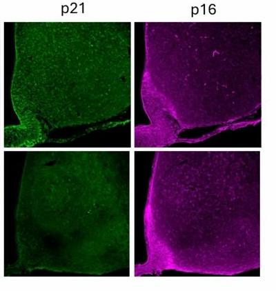
FH Bárbara (Verified Customer) (07-31-2025) | We used the p21 rabbit antibody from Proteintech for immunofluorescence on mouse brain, followed by confocal microscopy. The antibody produced a strong, specific signal . Minimal background and high signal-to-noise ratio made it easy to interpret.
 |

