- Phare
- Validé par KD/KO
Anticorps Polyclonal de lapin anti-DRG2
DRG2 Polyclonal Antibody for WB, IHC, IF/ICC, IP, ELISA
Hôte / Isotype
Lapin / IgG
Réactivité testée
Humain, rat, souris et plus (1)
Applications
WB, IHC, IF/ICC, IP, ELISA
Conjugaison
Non conjugué
N° de cat : 14743-1-AP
Synonymes
Galerie de données de validation
Applications testées
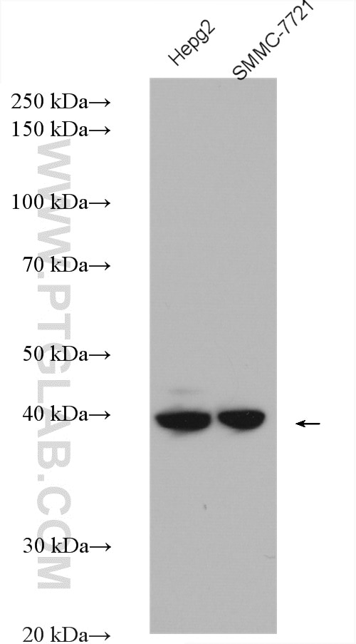
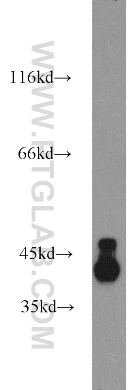
| Résultats positifs en WB | cellules HepG2, cellules SMMC-7721, tissu cérébral de souris |
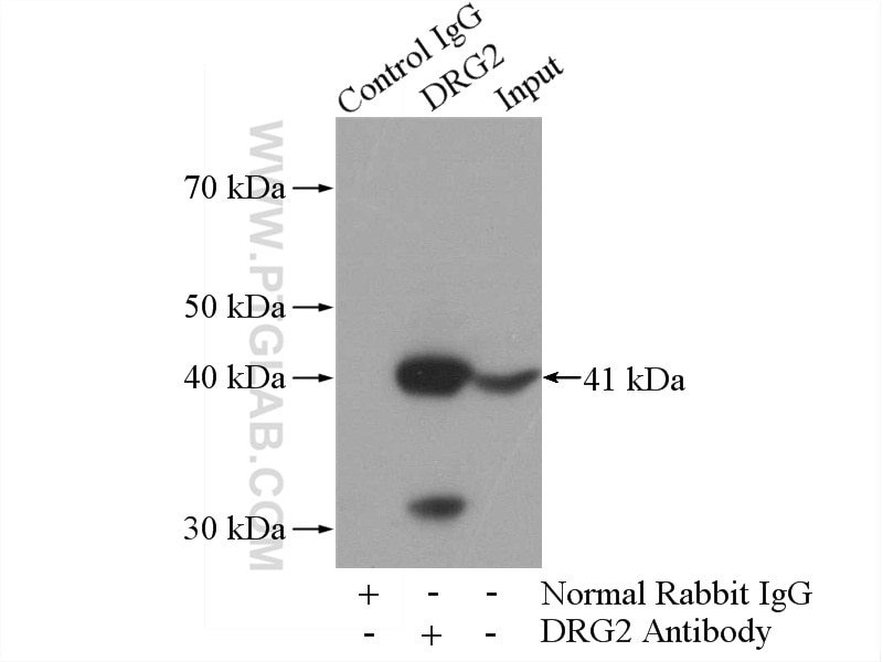
| Résultats positifs en IP | tissu cérébral de souris |
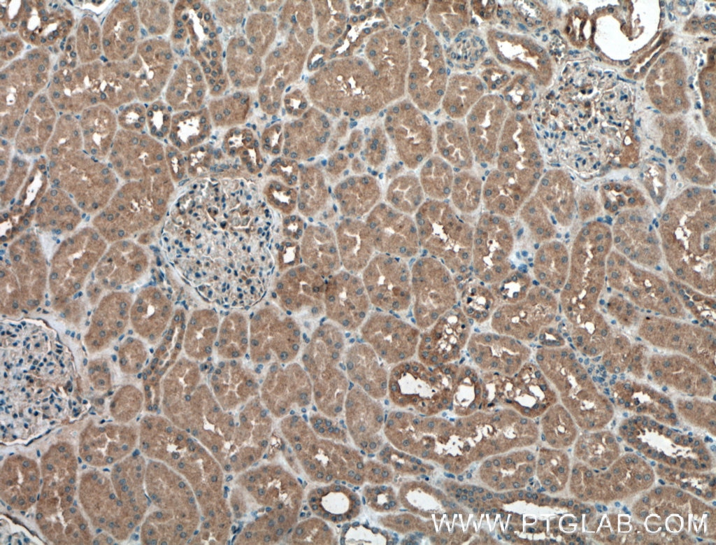
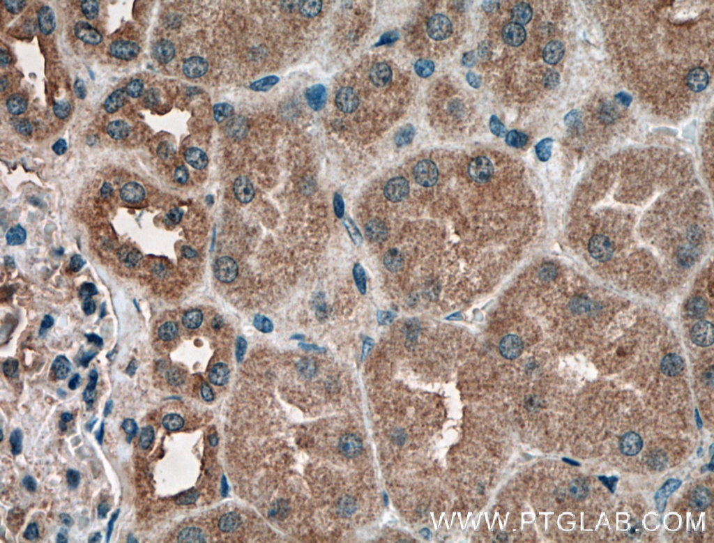
| Résultats positifs en IHC | tissu rénal humain, tissu cérébral de souris il est suggéré de démasquer l'antigène avec un tampon de TE buffer pH 9.0; (*) À défaut, 'le démasquage de l'antigène peut être 'effectué avec un tampon citrate pH 6,0. |
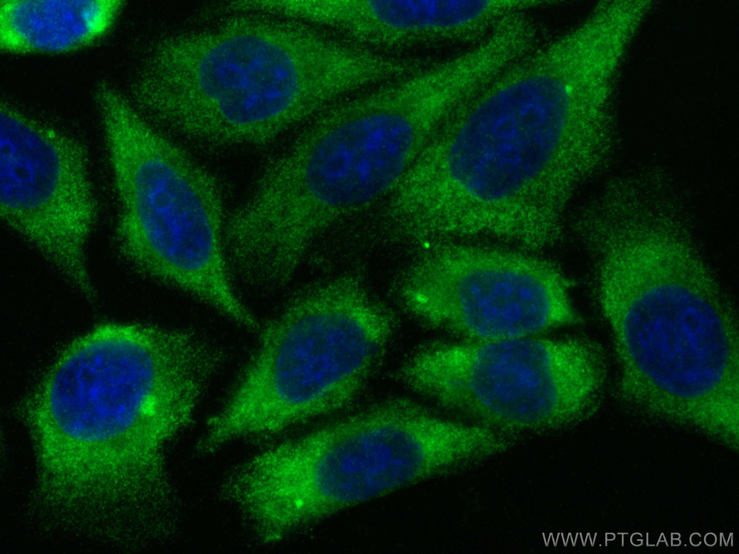
| Résultats positifs en IF/ICC | cellules HepG2, |
Dilution recommandée
| Application | Dilution |
|---|---|
| Western Blot (WB) | WB : 1:3000-1:8000 |
| Immunoprécipitation (IP) | IP : 0.5-4.0 ug for 1.0-3.0 mg of total protein lysate |
| Immunohistochimie (IHC) | IHC : 1:50-1:500 |
| Immunofluorescence (IF)/ICC | IF/ICC : 1:50-1:500 |
| It is recommended that this reagent should be titrated in each testing system to obtain optimal results. | |
| Sample-dependent, check data in validation data gallery | |
Applications publiées
| KD/KO | See 7 publications below |
| WB | See 15 publications below |
| IHC | See 1 publications below |
| IF | See 3 publications below |
| IP | See 1 publications below |
Informations sur le produit
14743-1-AP cible DRG2 dans les applications de WB, IHC, IF/ICC, IP, ELISA et montre une réactivité avec des échantillons Humain, rat, souris
| Réactivité | Humain, rat, souris |
| Réactivité citée | rat, Humain, singe, souris |
| Hôte / Isotype | Lapin / IgG |
| Clonalité | Polyclonal |
| Type | Anticorps |
| Immunogène | DRG2 Protéine recombinante Ag6433 |
| Nom complet | developmentally regulated GTP binding protein 2 |
| Masse moléculaire calculée | 41 kDa |
| Poids moléculaire observé | 41-45 kDa |
| Numéro d’acquisition GenBank | BC000493 |
| Symbole du gène | DRG2 |
| Identification du gène (NCBI) | 1819 |
| Conjugaison | Non conjugué |
| Forme | Liquide |
| Méthode de purification | Purification par affinité contre l'antigène |
| Tampon de stockage | PBS with 0.02% sodium azide and 50% glycerol |
| Conditions de stockage | Stocker à -20°C. Stable pendant un an après l'expédition. L'aliquotage n'est pas nécessaire pour le stockage à -20oC Les 20ul contiennent 0,1% de BSA. |
Informations générales
Developmentally regulated GTP-binding protein (DRG) is a new subfamily within the superfamily of GTP-binding proteins. Its expression is regulated during embryonic development. DRG2 plays a critical role in control of the cell cycle and apoptosis (PMID:27669826, PMID:14759600). DRG2 Regulates G2/M Progression via the Cyclin B1-Cdk1 Complex.
Protocole
| Product Specific Protocols | |
|---|---|
| WB protocol for DRG2 antibody 14743-1-AP | Download protocol |
| IHC protocol for DRG2 antibody 14743-1-AP | Download protocol |
| IF protocol for DRG2 antibody 14743-1-AP | Download protocol |
| IP protocol for DRG2 antibody 14743-1-AP | Download protocol |
| Standard Protocols | |
|---|---|
| Click here to view our Standard Protocols |
Publications
| Species | Application | Title |
|---|---|---|
Mol Ther Nucleic Acids Translatome and Transcriptome Profiling of Hypoxic-Induced Rat Cardiomyocytes. | ||
Int J Mol Sci DRG2 Depletion Promotes Endothelial Cell Senescence and Vascular Endothelial Dysfunction.
| ||
Oxid Med Cell Longev DRG2 Accelerates Senescence via Negative Regulation of SIRT1 in Human Diploid Fibroblasts
| ||
FASEB J Global analyses of mRNA expression in human sensory neurons reveal eIF5A as a conserved target for inflammatory pain. | ||
J Mol Med (Berl) Role of endosomal RANKL-LGR4 signaling during osteoclast differentiation
| ||
Mol Cell Endocrinol RWDD1 interacts with the ligand binding domain of the androgen receptor and acts as a coactivator of androgen-dependent transactivation. |
Avis
The reviews below have been submitted by verified Proteintech customers who received an incentive for providing their feedback.
FH Wojciech (Verified Customer) (09-12-2025) | The antibody against DRG2 protein showed a single band at ~40 kDa in T-cells, consistent with the expected molecular weight. The signal was clean with minimal background. No non-specific bands were observed.
|
