- Phare
- Validé par KD/KO
Anticorps Polyclonal de lapin anti-EDC4
EDC4 Polyclonal Antibody for WB, IHC, IF/ICC, ELISA
Hôte / Isotype
Lapin / IgG
Réactivité testée
Humain
Applications
WB, IHC, IF/ICC, ELISA
Conjugaison
Non conjugué
N° de cat : 17737-1-AP
Synonymes
Galerie de données de validation
 with sh-Control and sh-EDC4 transfected HeLa cells.")
 at dilution of 1:3000 incubated at room temperature for 1.5 hours.")
 at dilution of 1:600 (under 10x lens). Heat mediated antigen retrieval with Tris-EDTA buffer (pH 9.0).")
 at dilution of 1:600 (under 40x lens). Heat mediated antigen retrieval with Tris-EDTA buffer (pH 9.0).")
 at dilution of 1:600 (under 10x lens). Heat mediated antigen retrieval with Tris-EDTA buffer (pH 9.0).")
 at dilution of 1:600 (under 40x lens). Heat mediated antigen retrieval with Tris-EDTA buffer (pH 9.0).")
 fixed HeLa cells using EDC4 antibody (17737-1-AP) at dilution of 1:400 and CoraLite®488-Conjugated AffiniPure Goat Anti-Rabbit IgG(H+L), CL594-phalloidin (red).")
 fixed HeLa cells using EDC4 antibody (17737-1-AP) at dilution of 1:200 and CoraLite®488-Conjugated AffiniPure Goat Anti-Rabbit IgG(H+L), CL594-Phalloidin (red).")
Applications testées
| Résultats positifs en WB | cellules HeLa, cellules HepG2 |
| Résultats positifs en IHC | tissu de cancer du foie humain, tissu de cancer du côlon humain il est suggéré de démasquer l'antigène avec un tampon de TE buffer pH 9.0; (*) À défaut, 'le démasquage de l'antigène peut être 'effectué avec un tampon citrate pH 6,0. |
| Résultats positifs en IF/ICC | cellules HeLa, |
Dilution recommandée
| Application | Dilution |
|---|---|
| Western Blot (WB) | WB : 1:1000-1:6000 |
| Immunohistochimie (IHC) | IHC : 1:300-1:1200 |
| Immunofluorescence (IF)/ICC | IF/ICC : 1:200-1:800 |
| It is recommended that this reagent should be titrated in each testing system to obtain optimal results. | |
| Sample-dependent, check data in validation data gallery | |
Applications publiées
| KD/KO | See 1 publications below |
| WB | See 8 publications below |
| IF | See 9 publications below |
Informations sur le produit
17737-1-AP cible EDC4 dans les applications de WB, IHC, IF/ICC, ELISA et montre une réactivité avec des échantillons Humain
| Réactivité | Humain |
| Réactivité citée | Humain |
| Hôte / Isotype | Lapin / IgG |
| Clonalité | Polyclonal |
| Type | Anticorps |
| Immunogène | EDC4 Protéine recombinante Ag11784 |
| Nom complet | enhancer of mRNA decapping 4 |
| Masse moléculaire calculée | 1401 aa, 152 kDa |
| Poids moléculaire observé | 160 kDa |
| Numéro d’acquisition GenBank | BC064567 |
| Symbole du gène | EDC4 |
| Identification du gène (NCBI) | 23644 |
| Conjugaison | Non conjugué |
| Forme | Liquide |
| Méthode de purification | Purification par affinité contre l'antigène |
| Tampon de stockage | PBS with 0.02% sodium azide and 50% glycerol |
| Conditions de stockage | Stocker à -20°C. Stable pendant un an après l'expédition. L'aliquotage n'est pas nécessaire pour le stockage à -20oC Les 20ul contiennent 0,1% de BSA. |
Informations générales
Enhancer of mRNA-decapping protein 4 (EDC4) is a key regulator of mRNA decapping and degradation, playing a crucial role in the mRNA decay pathway. It is involved in the formation of processing bodies (P-bodies) in the cytoplasm, where it interacts with other decapping factors such as DCP1A and DCP2A to facilitate mRNA degradation (PMID: 16364915). EDC4 also interacts with the mammalian target of rapamycin complex 1 (mTORC1) and is involved in the regulation of immune system functions (PMID: 25514416; 25970328). Additionally, EDC4 has been shown to regulate DNA repair processes, with its deficiency leading to genome instability and hypersensitivity to DNA interstrand cross-linking drugs (PMID: 29511213).
Protocole
| Product Specific Protocols | |
|---|---|
| WB protocol for EDC4 antibody 17737-1-AP | Download protocol |
| IHC protocol for EDC4 antibody 17737-1-AP | Download protocol |
| IF protocol for EDC4 antibody 17737-1-AP | Download protocol |
| Standard Protocols | |
|---|---|
| Click here to view our Standard Protocols |
Publications
| Species | Application | Title |
|---|---|---|
Nat Cell Biol O-GlcNAcylation determines the translational regulation and phase separation of YTHDF proteins | ||
Cancer Res Pooled CRISPR Screening Identifies P-Bodies as Repressors of Cancer Epithelial-Mesenchymal Transition | ||
Acta Pharmacol Sin NSCLC cells sustain phase separation of cytoplasmic membrane-less organelles to protect themselves against cisplatin treatment | ||
Mol Ther Nucleic Acids The RNA binding protein QKI5 suppresses ovarian cancer via downregulating transcriptional coactivator TAZ. | ||
J Genet Genomics LSM14B coordinates protein component expression in the P-body and controls oocyte maturation | ||
FEBS Lett Insight into the function of the Golgi membrane protein GOLM1 in cholangiocytes through interactomic analysis |
Avis
The reviews below have been submitted by verified Proteintech customers who received an incentive for providing their feedback.
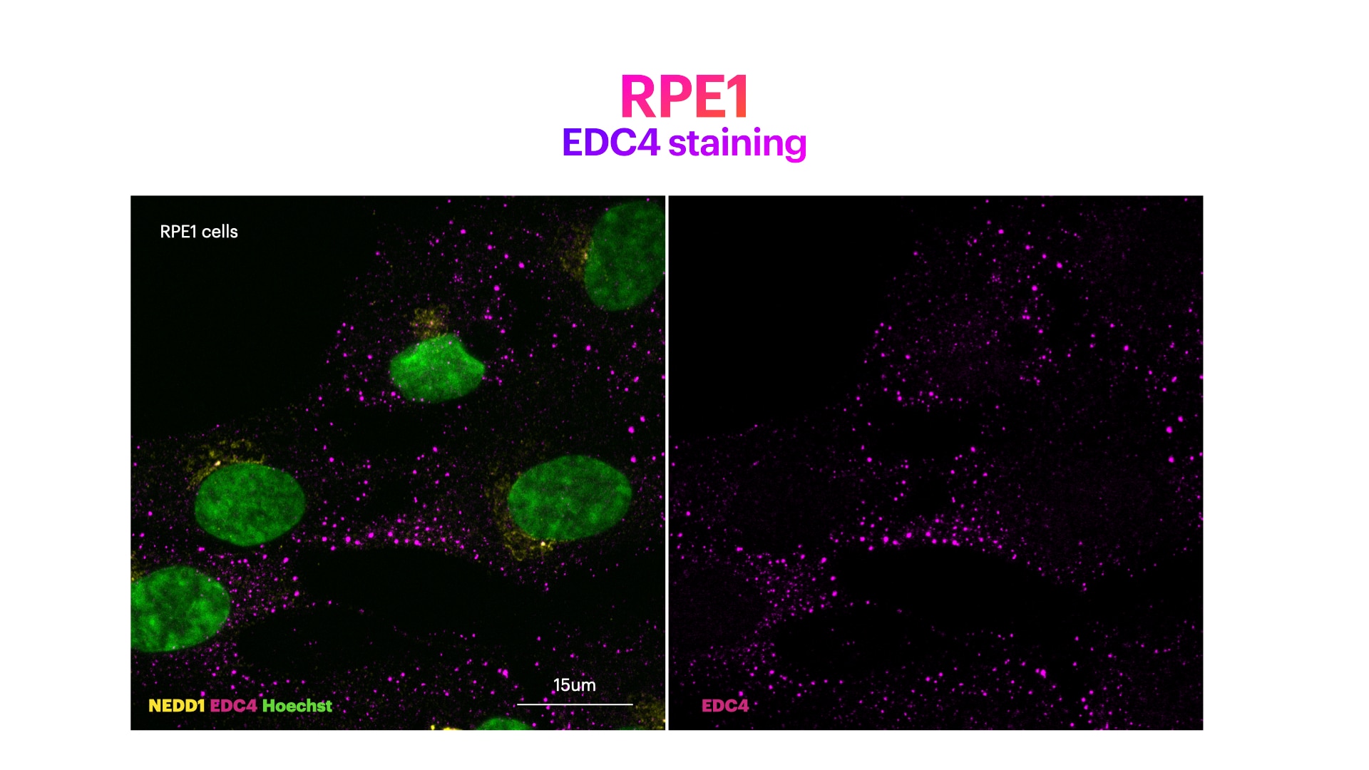
FH Elisa (Verified Customer) (03-01-2022) | RPE1 cells stained for Hoechst (DNA marker, in green), EDC4 (mRNA decapping protein 4 in (P)-bodies, in magenta) and NEDD1 (pericentriolar matrix marker, in yellow). RPE1 cells were fixed in 4%PFA for 15’. Cells were then washed with PBS. Membrane permeabilization was then performed with 0.3% Triton for 5'. Cells were finally incubated with blocking buffer (5% BSA+ 0.1% Tween in PBS) for 30' at RT. Primary antibody was diluted in blocking buffer 1:200 and incubated for 1h at room temperature. Alexa-555-Anti-rabbit was used as secondary antibody (1:600 dilution) (1h at room temperature).
 |

