- Phare
- Validé par KD/KO
Anticorps Polyclonal de lapin anti-EGFR-Specific
EGFR-Specific Polyclonal Antibody for WB, IP, ELISA
Hôte / Isotype
Lapin / IgG
Réactivité testée
Humain et plus (4)
Applications
WB, IHC, IF, IP, CoIP, ChIP, ELISA
Conjugaison
Non conjugué
N° de cat : 18986-1-AP
Synonymes
Galerie de données de validation
 at dilution of 1:3000 incubated at room temperature for 1.5 hours.")
 with sh-Control and sh-EGFR-Specific transfected HepG2 cells.")
 in Hela and REP1 cell by Dr.Kodani, Andrew.")
 at dilution of 1:6000 incubated at room temperature for 1.5 hours.")
 at dilution of 1:6000 incubated at room temperature for 1.5 hours.")
 at dilution of 1:1000 incubated at 4 degree celsius over night.")
 at dilution of 1:4000 incubated at room temperature for 1.5 hours.")
 at dilution of 1:500 incubated at room temperature for 1.5 hours.")
 with MCF-7 cells lysate 2500ug.")
Applications testées
| Résultats positifs en WB | cellules HepG2, cellules A431, cellules A549, cellules HeLa, cellules HeLa/RPE1, cellules L02, cellules MCF-7, cellules NCI-H1299, cellules NCL-H1299 |
| Résultats positifs en IP | cellules MCF-7 |
Dilution recommandée
| Application | Dilution |
|---|---|
| Western Blot (WB) | WB : 1:1000-1:6000 |
| Immunoprécipitation (IP) | IP : 0.5-4.0 ug for 1.0-3.0 mg of total protein lysate |
| It is recommended that this reagent should be titrated in each testing system to obtain optimal results. | |
| Sample-dependent, check data in validation data gallery | |
Informations sur le produit
18986-1-AP cible EGFR-Specific dans les applications de WB, IHC, IF, IP, CoIP, ChIP, ELISA et montre une réactivité avec des échantillons Humain
| Réactivité | Humain |
| Réactivité citée | rat, Chèvre, Humain, porc, souris |
| Hôte / Isotype | Lapin / IgG |
| Clonalité | Polyclonal |
| Type | Anticorps |
| Immunogène | Peptide |
| Nom complet | epidermal growth factor receptor (erythroblastic leukemia viral (v-erb-b) oncogene homolog, avian) |
| Masse moléculaire calculée | 134 kDa |
| Poids moléculaire observé | 145-165 kDa |
| Numéro d’acquisition GenBank | NM_005228 |
| Symbole du gène | EGFR |
| Identification du gène (NCBI) | 1956 |
| Conjugaison | Non conjugué |
| Forme | Liquide |
| Méthode de purification | Purification par affinité contre l'antigène |
| Tampon de stockage | PBS with 0.02% sodium azide and 50% glycerol |
| Conditions de stockage | Stocker à -20°C. Stable pendant un an après l'expédition. L'aliquotage n'est pas nécessaire pour le stockage à -20oC Les 20ul contiennent 0,1% de BSA. |
Informations générales
ERBB1, also named as EGFR, is a receptor for EGF, but also for other members of the EGF family, as TGF-alpha, amphiregulin, betacellulin, heparin-binding EGF-like growth factor, GP30 and vaccinia virus growth factor. ERBB1 is involved in the control of cell growth and differentiation. Phosphorylates MUC1 in breast cancer cells and increases the interaction of MUC1 with C-SRC and CTNNB1/beta-catenin. Isoform 2/truncated isoform of ERBB1 may act as an antagonist. EGFR is associated with lung cancer. The antibody is specific to isoform1.
Protocole
| Product Specific Protocols | |
|---|---|
| WB protocol for EGFR-Specific antibody 18986-1-AP | Download protocol |
| IP protocol for EGFR-Specific antibody 18986-1-AP | Download protocol |
| Standard Protocols | |
|---|---|
| Click here to view our Standard Protocols |
Publications
| Species | Application | Title |
|---|---|---|
Nat Microbiol Interferon-induced transmembrane protein-1 competitively blocks Ephrin receptor A2-mediated Epstein-Barr virus entry into epithelial cells | ||
Cell Res In vivo self-assembled small RNAs as a new generation of RNAi therapeutics.
| ||
Nat Commun Inhalable liposomal delivery of osimertinib and DNA for treating primary and metastasis lung cancer | ||
Cancer Commun (Lond) Blockage of EGFR/AKT and mevalonate pathways synergize the antitumor effect of temozolomide by reprogramming energy metabolism in glioblastoma | ||
Biosens Bioelectron Microneedle patches integrated with lateral flow cassettes for blood-free chronic kidney disease point-of-care testing during a pandemic. |
Avis
The reviews below have been submitted by verified Proteintech customers who received an incentive for providing their feedback.
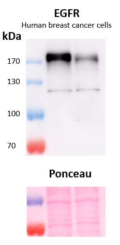
FH Marina (Verified Customer) (02-13-2023) | A stronger band can be observed slighly above 170 kDa and a fainter band below 130 kDa. Ponceau red is used as total protein loading control. Primary antibody dilution 1:500, overnight incubation at 4ºC. Exposure time 3 minutes.
 |
FH Clarisse (Verified Customer) (12-12-2022) | This antibody works very well on tissue fixed overnight at 4 degrees C with 2% PFA. No antigen retrieval required.
|
FH Morgan (Verified Customer) (10-26-2021) | I had luck with this antibody as seen from the image attached. It took a little bit of time to develop though. Maybe using more than 1:500 would be better. Unfortunately I couldn't get the phosphorylated version (p-EGF) to work though. I used OV90 (ovarian cancer) cells
 |
FH K (Verified Customer) (12-20-2020) | worked well for WB (1:1000)IF (1:200)and IHC (1:200)
 |
FH XB (Verified Customer) (10-29-2020) | It is OK to use it to probe EGFR (about 180 kD), but there are some unspecific bands at the lower part of the gel.
 |

