- Phare
- Validé par KD/KO
Anticorps Polyclonal de lapin anti-EHD1
EHD1 Polyclonal Antibody for WB, IHC, IP, ELISA
Hôte / Isotype
Lapin / IgG
Réactivité testée
Humain, rat, souris
Applications
WB, IHC, IF, IP, ELISA
Conjugaison
Non conjugué
N° de cat : 24657-1-AP
Synonymes
Galerie de données de validation
 at dilution of 1:2000 incubated at room temperature for 1.5 hours.")
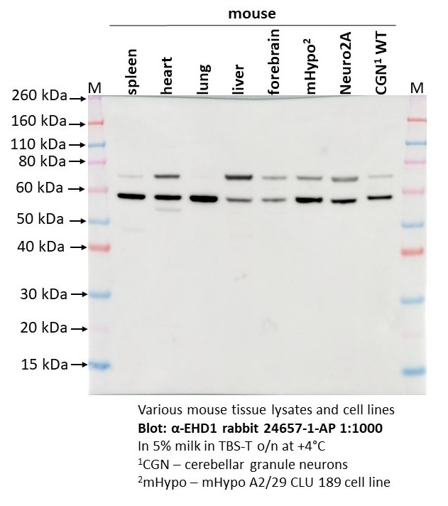
 at dilution of 1:1000 incubated at room temperature for 1.5 hours.")
 at dilution of 1:1000 incubated at room temperature for 1.5 hours.")
 at dilution of 1:1000 incubated at room temperature for 1.5 hours.")
 with mouse testis tissue lysate 4000 ug.")
 at dilution of 1:200 (under 20x lens). Heat mediated antigen retrieval with Tris-EDTA buffer (pH 9.0).")
 at dilution of 1:200 (under 20x lens). Heat mediated antigen retrieval with Tris-EDTA buffer (pH 9.0).")
 at dilution of 1:200 (under 20x lens). Heat mediated antigen retrieval with Tris-EDTA buffer (pH 9.0).")
 at dilution of 1:200 (under 20x lens). Heat mediated antigen retrieval with Tris-EDTA buffer (pH 9.0).")
Applications testées
| Résultats positifs en WB | tissu pulmonaire de souris, cellules HeLa, tissu cérébral de souris, tissu pulmonaire de rat, tissu testiculaire de souris |
| Résultats positifs en IP | tissu testiculaire de souris, |
| Résultats positifs en IHC | tissu de cancer de l'estomac humain, human intrahepatic cholangiocarcinoma tissue il est suggéré de démasquer l'antigène avec un tampon de TE buffer pH 9.0; (*) À défaut, 'le démasquage de l'antigène peut être 'effectué avec un tampon citrate pH 6,0. |
Dilution recommandée
| Application | Dilution |
|---|---|
| Western Blot (WB) | WB : 1:1000-1:4000 |
| Immunoprécipitation (IP) | IP : 0.5-4.0 ug for 1.0-3.0 mg of total protein lysate |
| Immunohistochimie (IHC) | IHC : 1:50-1:500 |
| It is recommended that this reagent should be titrated in each testing system to obtain optimal results. | |
| Sample-dependent, check data in validation data gallery | |
Applications publiées
| KD/KO | See 3 publications below |
| WB | See 4 publications below |
| IHC | See 1 publications below |
| IF | See 2 publications below |
Informations sur le produit
24657-1-AP cible EHD1 dans les applications de WB, IHC, IF, IP, ELISA et montre une réactivité avec des échantillons Humain, rat, souris
| Réactivité | Humain, rat, souris |
| Réactivité citée | Humain, souris |
| Hôte / Isotype | Lapin / IgG |
| Clonalité | Polyclonal |
| Type | Anticorps |
| Immunogène | EHD1 Protéine recombinante Ag18400 |
| Nom complet | EH-domain containing 1 |
| Masse moléculaire calculée | 534 aa, 61 kDa |
| Poids moléculaire observé | 61 kDa |
| Numéro d’acquisition GenBank | BC104799 |
| Symbole du gène | EHD1 |
| Identification du gène (NCBI) | 10938 |
| Conjugaison | Non conjugué |
| Forme | Liquide |
| Méthode de purification | Purification par affinité contre l'antigène |
| Tampon de stockage | PBS with 0.02% sodium azide and 50% glycerol |
| Conditions de stockage | Stocker à -20°C. Stable pendant un an après l'expédition. L'aliquotage n'est pas nécessaire pour le stockage à -20oC Les 20ul contiennent 0,1% de BSA. |
Informations générales
EHD1 (Eps15 Homology Domain Containing 1) is a protein that plays a crucial role in endocytic recycling, which is the process by which cells recycle their membrane components. It is one of four paralogs in mammals (EHD1-4) and is involved in several membrane trafficking pathways. EHD1 is particularly well-studied and is known to regulate the recycling of various cell surface receptors back to the cell surface after they have been endocytosed. This process is essential for maintaining the proper distribution of receptors and other membrane proteins, which in turn affects cellular signaling and function. EHD1 has been implicated in the regulation of the transferrin receptor (TfR), major histocompatibility complex (MHC) class I proteins, β1 integrins, and other receptors. It has also been shown to interact with Rab11-FIP2 and is localized to peripheral endosomes, suggesting a role in the transport of receptors from early endosomes to the endocytic recycling compartment (ERC). Furthermore, EHD1 has been linked to dynein motors that drive transport from early endosomes to the ERC via a complex including MICAL-L1 and the collapsin response mediator protein-2 (Crmp2).
Protocole
| Product Specific Protocols | |
|---|---|
| WB protocol for EHD1 antibody 24657-1-AP | Download protocol |
| IHC protocol for EHD1 antibody 24657-1-AP | Download protocol |
| IP protocol for EHD1 antibody 24657-1-AP | Download protocol |
| Standard Protocols | |
|---|---|
| Click here to view our Standard Protocols |
Publications
| Species | Application | Title |
|---|---|---|
EMBO J Insufficiency of ciliary cholesterol in hereditary Zellweger syndrome.
| ||
Sci Rep Rab35 and its effectors promote formation of tunneling nanotubes in neuronal cells.
| ||
Biochem Biophys Res Commun A novel CDK-independent function of p27Kip1 in preciliary vesicle trafficking during ciliogenesis. | ||
J Extracell Vesicles Breast Cancer-Derived Extracellular Vesicles Modulate the Cytoplasmic and Cytoskeletal Dynamics of Blood-Brain Barrier Endothelial Cells |
Avis
The reviews below have been submitted by verified Proteintech customers who received an incentive for providing their feedback.
FH Olga (Verified Customer) (09-12-2025) | The blot is quite clear but a bit confusing that there are two bands in mouse cell lines and tissues.
 |
FH Kyosuke (Verified Customer) (06-12-2019) | I am working on thrombosis study. I use this for mouse platelet Western blot and it works very well.
|
FH Juan (Verified Customer) (05-02-2019) | It works well with a single band
|

