- Phare
- Validé par KD/KO
Anticorps Polyclonal de lapin anti-ENDOG
ENDOG Polyclonal Antibody for WB, IHC, IP, ELISA
Hôte / Isotype
Lapin / IgG
Réactivité testée
Humain, rat, souris
Applications
WB, IHC, IF, IP, CoIP, ELISA
Conjugaison
Non conjugué
N° de cat : 22148-1-AP
Synonymes
Galerie de données de validation
 at dilution of 1:8000 incubated at room temperature for 1.5 hours.")
 at dilution of 1:1000 incubated at room temperature for 1.5 hours.")
 at dilution of 1:1000 incubated at room temperature for 1.5 hours.")
 at dilution of 1:1000 incubated at room temperature for 1.5 hours.")
 at dilution of 1:500 incubated at room temperature for 1.5 hours.")
 at dilution of 1:1000 incubated at room temperature for 1.5 hours.")
 at dilution of 1:1000 incubated at 4 degree celsius over night.")
 with mouse heart tissue lysate 4000ug.")
 at dilution of 1:800 (under 20x lens). Heat mediated antigen retrieval with Tris-EDTA buffer (pH 9.0).")
Applications testées
| Résultats positifs en WB | tissu cardiaque de souris, tissu de muscle squelettique de souris, tissu hépatique de rat, tissu hépatique de souris, tissu rénal de rat, tissu rénal de souris |
| Résultats positifs en IP | tissu cardiaque de souris |
| Résultats positifs en IHC | human ovary cancer tissue, il est suggéré de démasquer l'antigène avec un tampon de TE buffer pH 9.0; (*) À défaut, 'le démasquage de l'antigène peut être 'effectué avec un tampon citrate pH 6,0. |
Dilution recommandée
| Application | Dilution |
|---|---|
| Western Blot (WB) | WB : 1:2000-1:16000 |
| Immunoprécipitation (IP) | IP : 0.5-4.0 ug for 1.0-3.0 mg of total protein lysate |
| Immunohistochimie (IHC) | IHC : 1:400-1:1600 |
| It is recommended that this reagent should be titrated in each testing system to obtain optimal results. | |
| Sample-dependent, check data in validation data gallery | |
Applications publiées
| KD/KO | See 1 publications below |
| WB | See 12 publications below |
| IHC | See 1 publications below |
| IF | See 1 publications below |
| CoIP | See 1 publications below |
Informations sur le produit
22148-1-AP cible ENDOG dans les applications de WB, IHC, IF, IP, CoIP, ELISA et montre une réactivité avec des échantillons Humain, rat, souris
| Réactivité | Humain, rat, souris |
| Réactivité citée | rat, Humain, souris |
| Hôte / Isotype | Lapin / IgG |
| Clonalité | Polyclonal |
| Type | Anticorps |
| Immunogène | ENDOG Protéine recombinante Ag17739 |
| Nom complet | endonuclease G |
| Masse moléculaire calculée | 297 aa, 33 kDa |
| Poids moléculaire observé | 27-30 kDa |
| Numéro d’acquisition GenBank | BC016351 |
| Symbole du gène | ENDOG |
| Identification du gène (NCBI) | 2021 |
| Conjugaison | Non conjugué |
| Forme | Liquide |
| Méthode de purification | Purification par affinité contre l'antigène |
| Tampon de stockage | PBS with 0.02% sodium azide and 50% glycerol |
| Conditions de stockage | Stocker à -20°C. Stable pendant un an après l'expédition. L'aliquotage n'est pas nécessaire pour le stockage à -20oC Les 20ul contiennent 0,1% de BSA. |
Informations générales
Endonuclease G, also named as EndoG, is a mitochondrial protein. It's a nuclease which was first characterized in bovine heart mitochondrial extracts. It's involved in many cellular process, including apoptosis, paternal mitochondrial elimination and autophage (PMID:33473107). It is a nuclear encoded, sugar-non-specific (PMID:15066427) and well-conserved nuclease (PMID:17244531). It can be released from the mitochondria and translocated to the nucleus where it induces fragmentation of DNA, leading to apoptosis (PMID:11452314). EndoG is a 297-amino-acid long protein with a molecular weight of 30-35 kDa. There is a homodimer form with MW about 60-70 kDa.
Protocole
| Product Specific Protocols | |
|---|---|
| WB protocol for ENDOG antibody 22148-1-AP | Download protocol |
| IHC protocol for ENDOG antibody 22148-1-AP | Download protocol |
| IP protocol for ENDOG antibody 22148-1-AP | Download protocol |
| Standard Protocols | |
|---|---|
| Click here to view our Standard Protocols |
Publications
| Species | Application | Title |
|---|---|---|
Chem Biol Interact O-Alkylated derivatives of quercetin induce apoptosis of MCF-7 cells via a caspase-independent mitochondrial pathway. | ||
Inflammation Chlorogenic Acid Alleviates Hepatic Ischemia-Reperfusion Injury by Inhibiting Oxidative Stress, Inflammation, and Mitochondria-Mediated Apoptosis In Vivo and In Vitro | ||
Front Pharmacol Quercitrin Attenuates Acetaminophen-Induced Acute Liver Injury by Maintaining Mitochondrial Complex I Activity. | ||
Front Pharmacol Emodin Induced SREBP1-Dependent and SREBP1-Independent Apoptosis in Hepatocellular Carcinoma Cells. | ||
Metallomics Induction of mitochondrial apoptosis pathway mediated through caspase-8 and c-Jun N-terminal kinase by cadmium-activated Fas in rat cortical neurons. | ||
Int J Mol Sci Proteomics Analysis of Tangeretin-Induced Apoptosis through Mitochondrial Dysfunction in Bladder Cancer Cells. |
Avis
The reviews below have been submitted by verified Proteintech customers who received an incentive for providing their feedback.
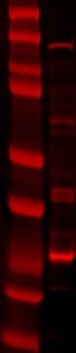
FH Lana (Verified Customer) (06-11-2021) | SDS-PAGE: 40 ug/ul RIPA protein lysate, 4-12% Bis-Tris gradient gel. Transfer: Immobilon-FL transfer membranes (Millipore) O/N at 30V, 4C. Blocking: SEA Block Blocking Buffer 1h, room T. Primary Ab: O/N incubation at 4C, 1:1000. Secondary Ab: IRDye 680LT Goat anti-Rabbit, 1:15000. Lines of WB image: 1 – protein ladder, 2 – mitochondria fraction lysate.
 |

