- Phare
- Validé par KD/KO
Anticorps Polyclonal de lapin anti-ERG
ERG Polyclonal Antibody for WB, IHC, IF/ICC, IP, ELISA
Hôte / Isotype
Lapin / IgG
Réactivité testée
Humain, rat, souris
Applications
WB, IHC, IF/ICC, IP, ELISA
Conjugaison
Non conjugué
N° de cat : 14356-1-AP
Synonymes
Galerie de données de validation
Applications testées
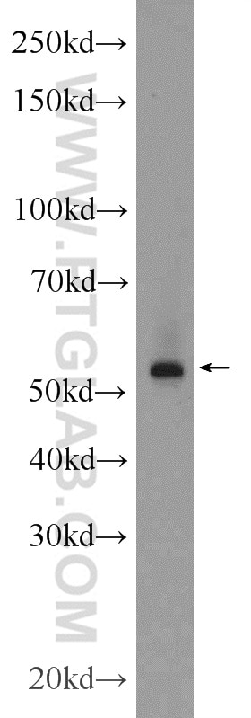
| Résultats positifs en WB | cellules MCF-7, |
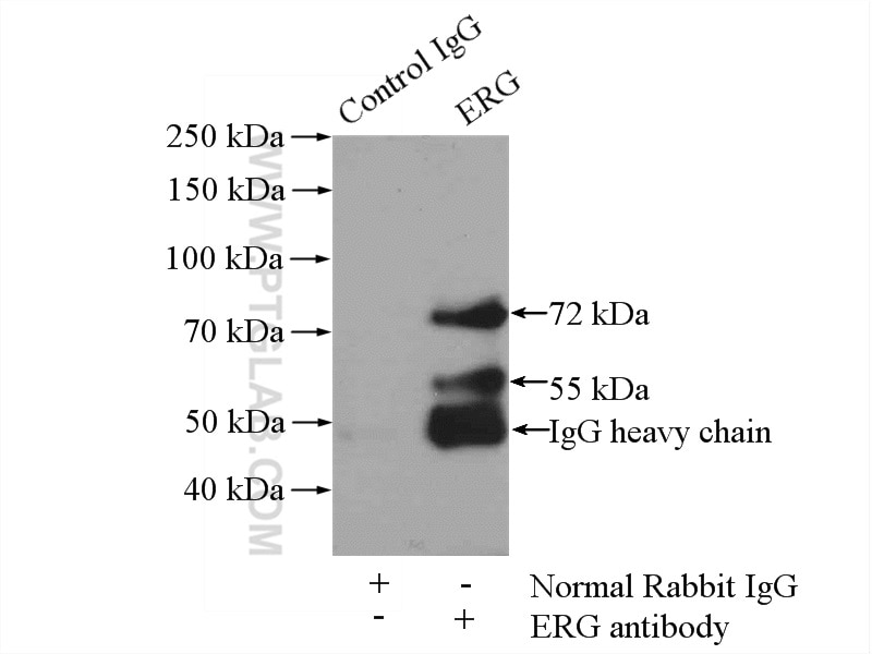
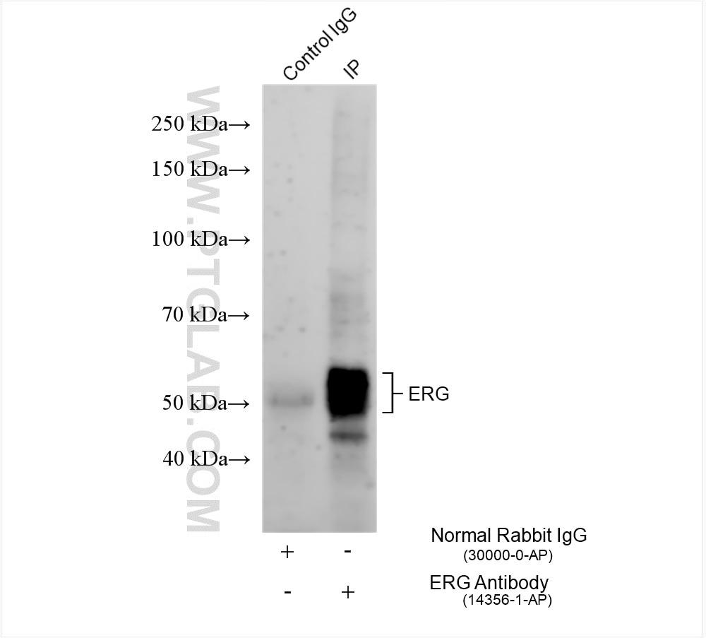
| Résultats positifs en IP | cellules MCF-7, tissu cardiaque de rat |
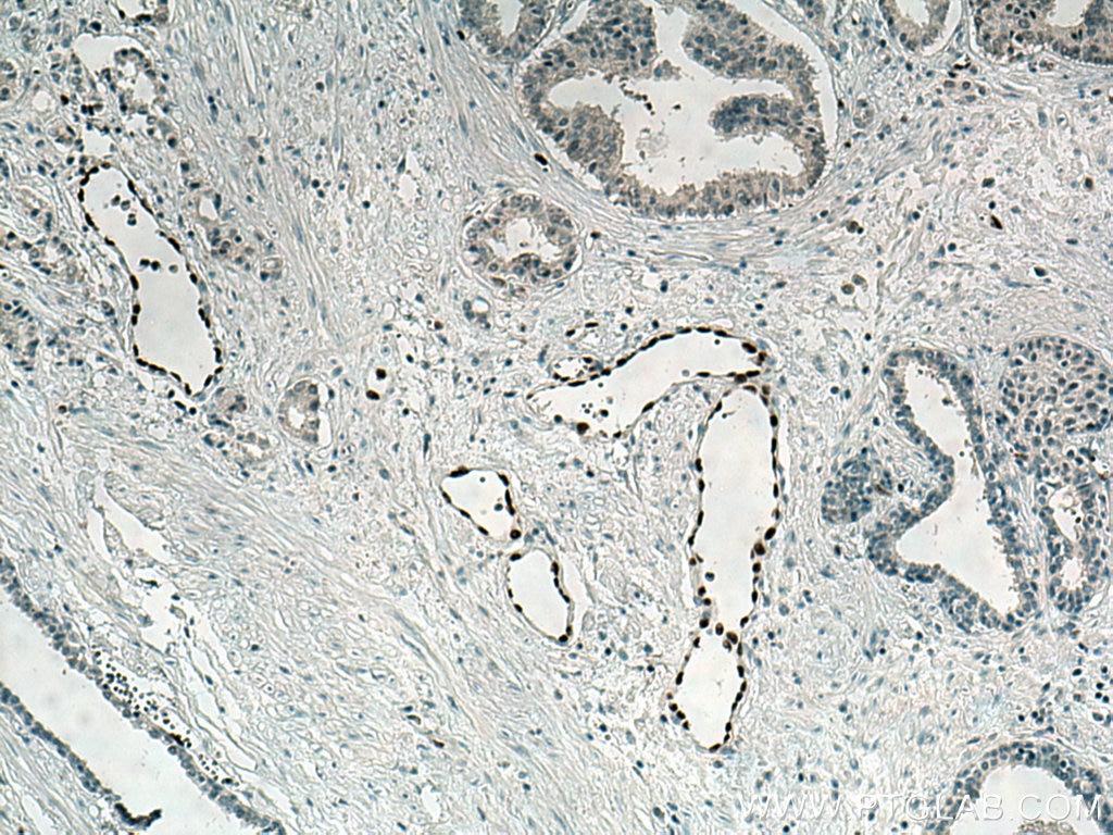
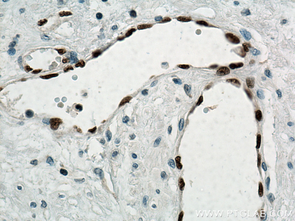
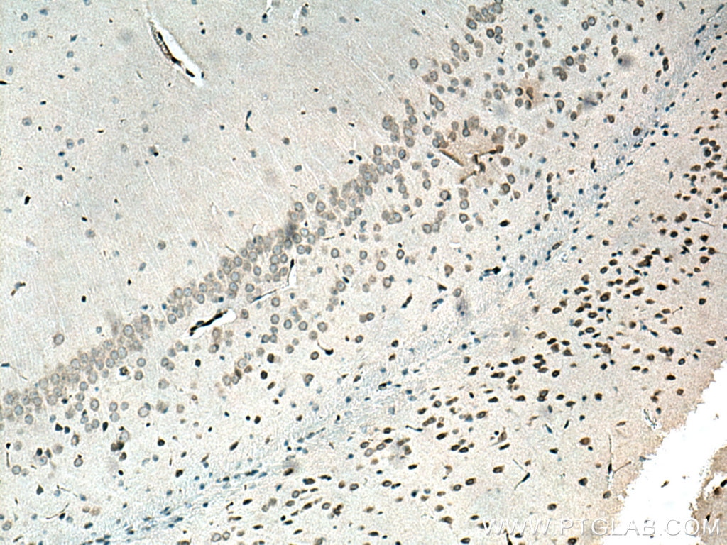
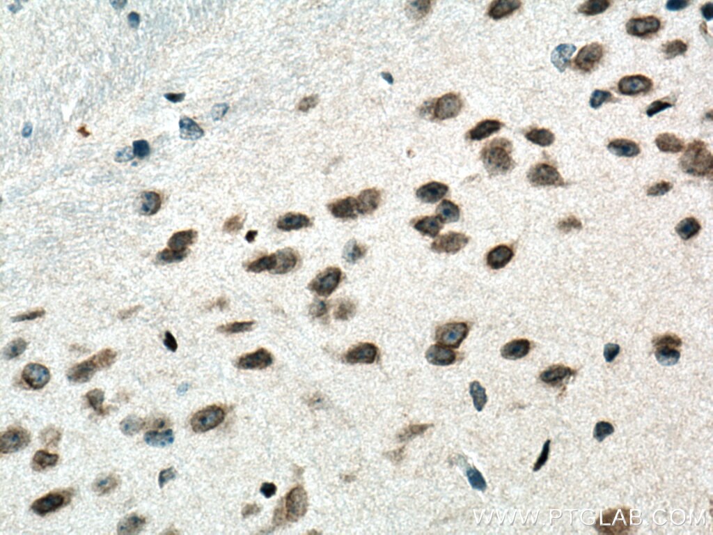
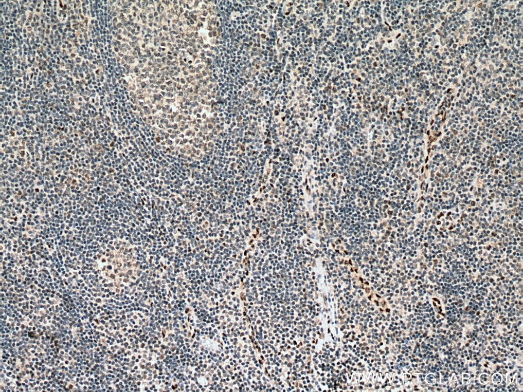
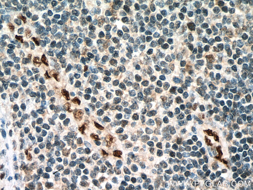
| Résultats positifs en IHC | tissu de cancer de la prostate humain, tissu cérébral de souris, tissu d'amygdalite humain il est suggéré de démasquer l'antigène avec un tampon de TE buffer pH 9.0; (*) À défaut, 'le démasquage de l'antigène peut être 'effectué avec un tampon citrate pH 6,0. |
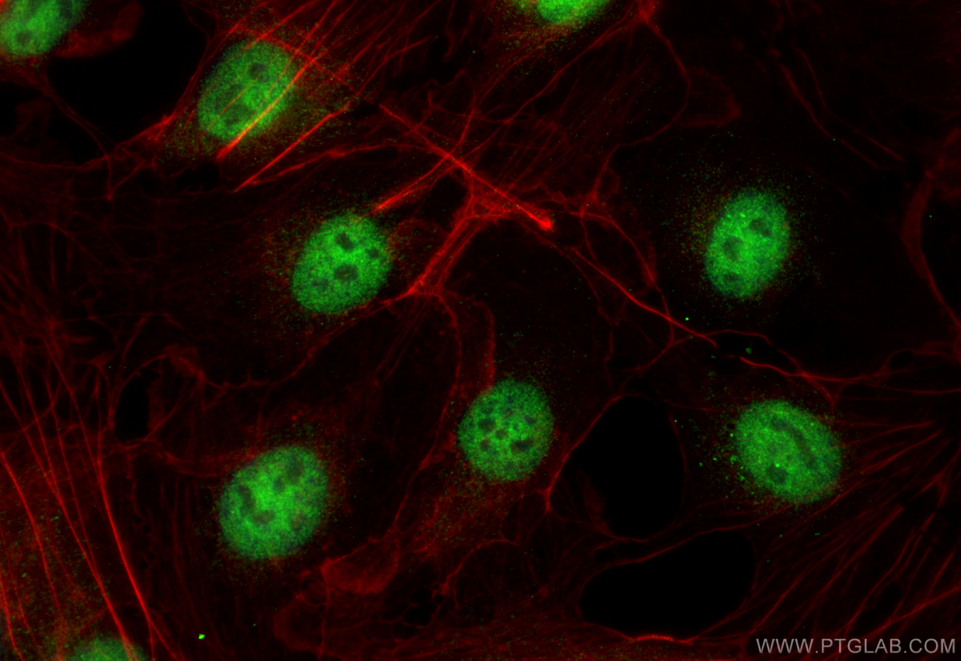
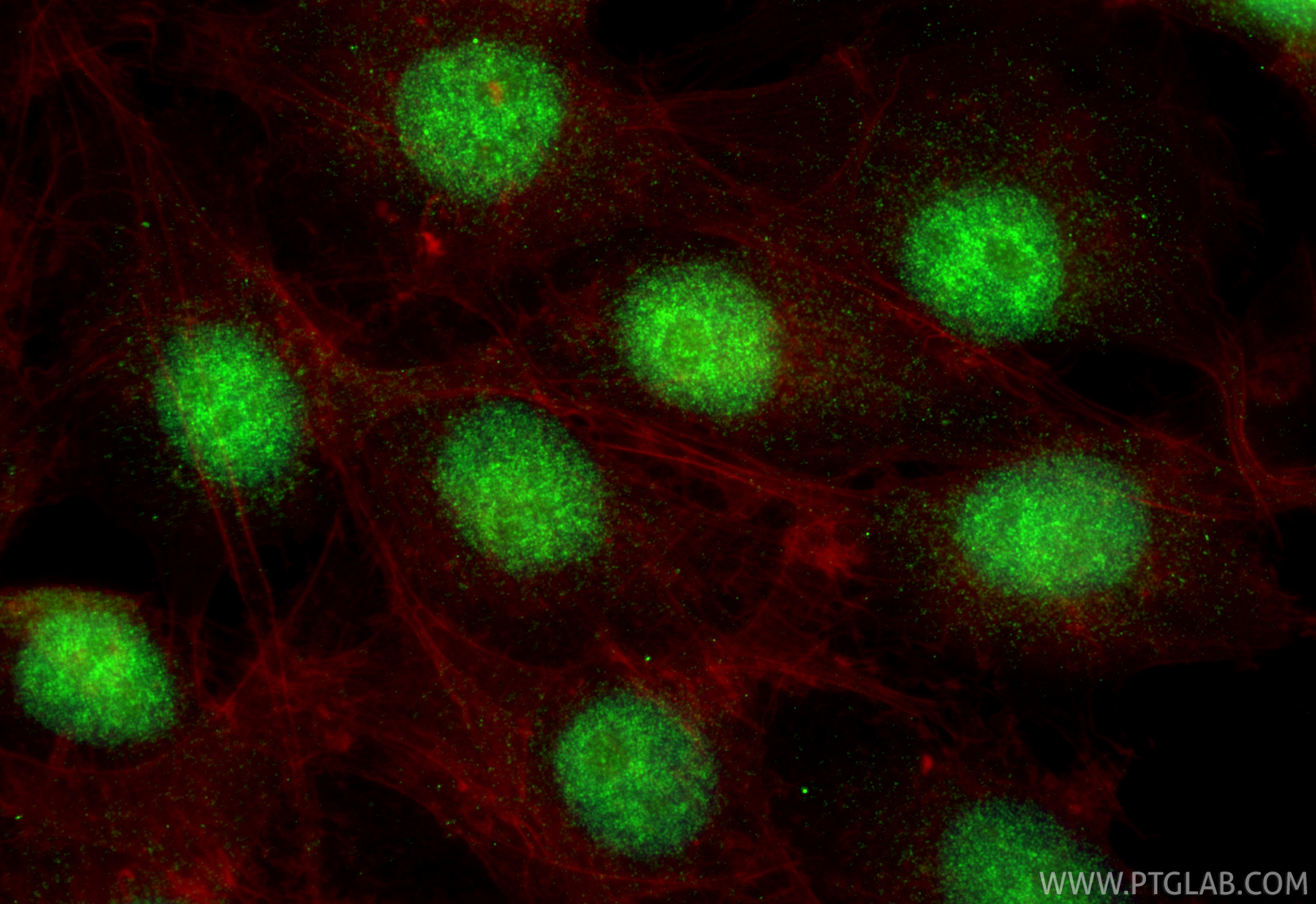
| Résultats positifs en IF/ICC | MG-63 cells, cellules HUVEC |
Dilution recommandée
| Application | Dilution |
|---|---|
| Western Blot (WB) | WB : 1:500-1:1000 |
| Immunoprécipitation (IP) | IP : 0.5-4.0 ug for 1.0-3.0 mg of total protein lysate |
| Immunohistochimie (IHC) | IHC : 1:600-1:2400 |
| Immunofluorescence (IF)/ICC | IF/ICC : 1:50-1:500 |
| It is recommended that this reagent should be titrated in each testing system to obtain optimal results. | |
| Sample-dependent, check data in validation data gallery | |
Applications publiées
| KD/KO | See 1 publications below |
| WB | See 5 publications below |
| IHC | See 2 publications below |
| IF | See 2 publications below |
Informations sur le produit
14356-1-AP cible ERG dans les applications de WB, IHC, IF/ICC, IP, ELISA et montre une réactivité avec des échantillons Humain, rat, souris
| Réactivité | Humain, rat, souris |
| Réactivité citée | Humain, souris |
| Hôte / Isotype | Lapin / IgG |
| Clonalité | Polyclonal |
| Type | Anticorps |
| Immunogène | ERG Protéine recombinante Ag5708 |
| Nom complet | v-ets erythroblastosis virus E26 oncogene homolog (avian) |
| Masse moléculaire calculée | 55 kDa |
| Poids moléculaire observé | 55 kDa |
| Numéro d’acquisition GenBank | BC040168 |
| Symbole du gène | ERG |
| Identification du gène (NCBI) | 2078 |
| Conjugaison | Non conjugué |
| Forme | Liquide |
| Méthode de purification | Purification par affinité contre l'antigène |
| Tampon de stockage | PBS with 0.02% sodium azide and 50% glycerol |
| Conditions de stockage | Stocker à -20°C. Stable pendant un an après l'expédition. L'aliquotage n'est pas nécessaire pour le stockage à -20oC Les 20ul contiennent 0,1% de BSA. |
Informations générales
ERG (ETS-related gene) is a member of the ETS family of transcription factors, which are downstream effectors of mitogenic signaling transduction pathway. ERG is a key regulator of cell proliferation, differentiation, angiogenesis, inflammation, and apoptosis. ERG is involved in chromosomal translocations, resulting in different fusion gene products, such as TMPSSR2-ERG and NDRG1-ERG in prostate cancer, EWS-ERG in Ewing's sarcoma and FUS-ERG in acute myeloid leukemia.
Protocole
| Product Specific Protocols | |
|---|---|
| WB protocol for ERG antibody 14356-1-AP | Download protocol |
| IHC protocol for ERG antibody 14356-1-AP | Download protocol |
| IF protocol for ERG antibody 14356-1-AP | Download protocol |
| IP protocol for ERG antibody 14356-1-AP | Download protocol |
| Standard Protocols | |
|---|---|
| Click here to view our Standard Protocols |
Publications
| Species | Application | Title |
|---|---|---|
Cell Stem Cell PSPC1 exerts an oncogenic role in AML by regulating a leukemic transcription program in cooperation with PU.1 | ||
iScience Resident vascular Sca1+ progenitors differentiate into endothelial cells in vascular remodeling via miR-145-5p/ERG signaling pathway | ||
Cell Biosci Profile of chimeric RNAs and TMPRSS2-ERG e2e4 isoform in neuroendocrine prostate cancer | ||
J Cancer miR-223-5p targeting ERG inhibits prostate cancer cell proliferation and migration.
| ||
Cell Death Discov HDAC11 promotes both NLRP3/caspase-1/GSDMD and caspase-3/GSDME pathways causing pyroptosis via ERG in vascular endothelial cells. | ||
Exp Ther Med Rare angiolymphoid hyperplasia with eosinophilia examined through fine needle aspiration cytology, histopathology and immunophenotypic characterization: A case report |
