- Phare
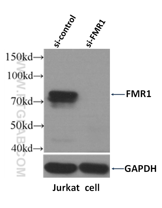
- Validé par KD/KO
Anticorps Polyclonal de lapin anti-FMR1
FMR1 Polyclonal Antibody for WB, IHC, IF/ICC, FC (Intra), IP, ELISA
Hôte / Isotype
Lapin / IgG
Réactivité testée
Humain, souris
Applications
WB, IHC, IF/ICC, FC (Intra), IP, ELISA
Conjugaison
Non conjugué
N° de cat : 13755-1-AP
Synonymes
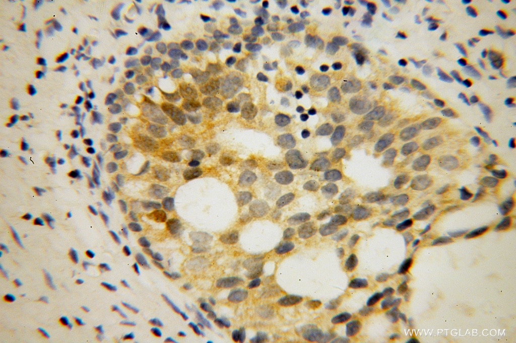
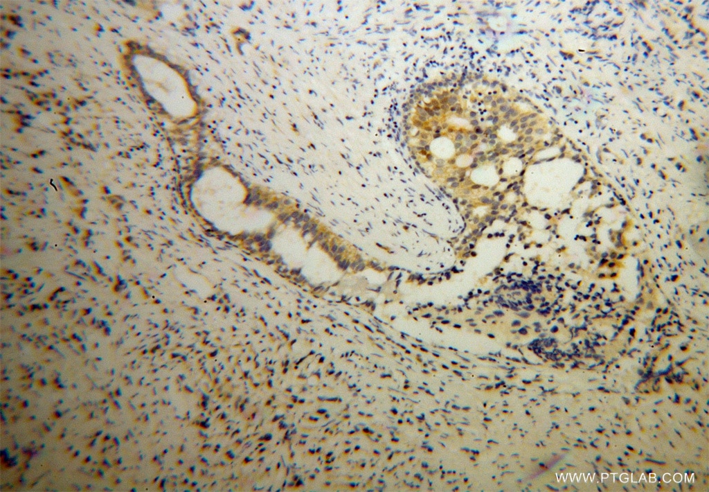
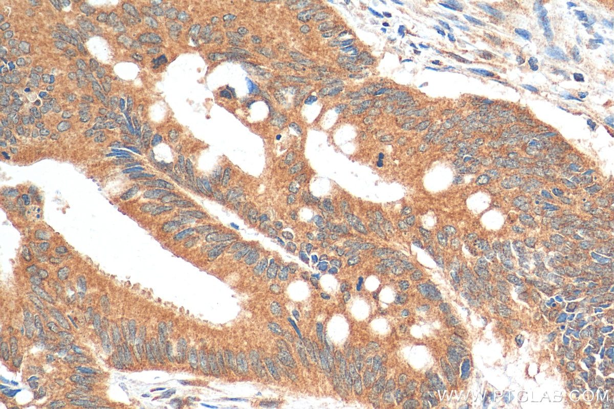
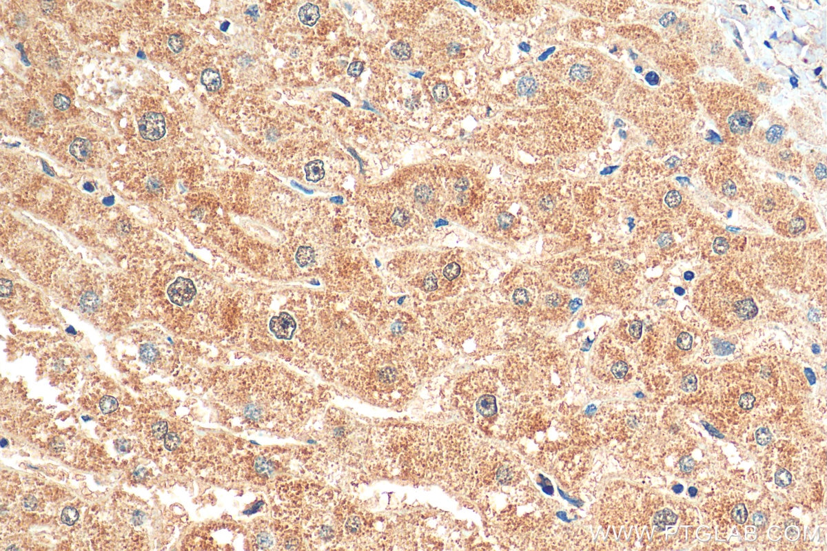
Galerie de données de validation
Applications testées
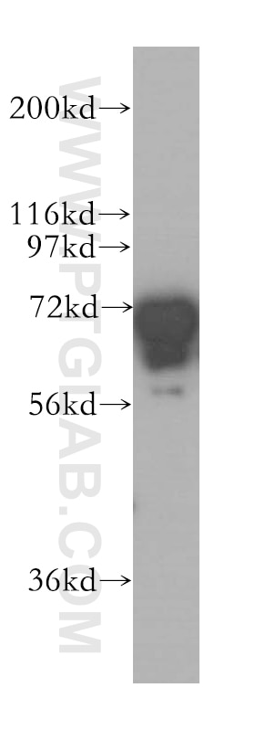
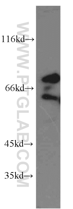
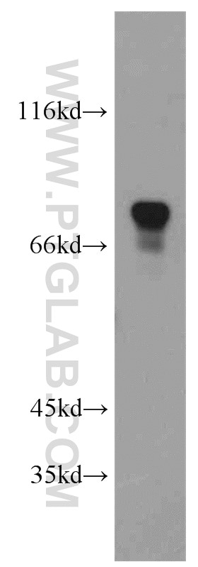
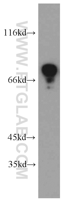
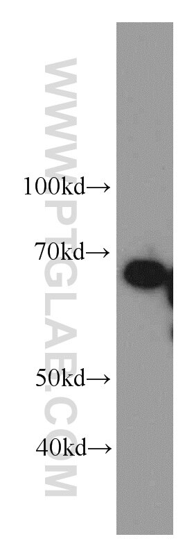
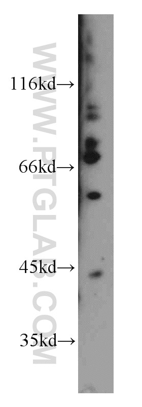
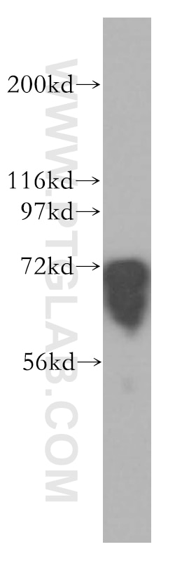
| Résultats positifs en WB | cellules K-562, cellules HEK-293, cellules HeLa, cellules Jurkat, tissu cérébral de souris, tissu de thymus de souris |
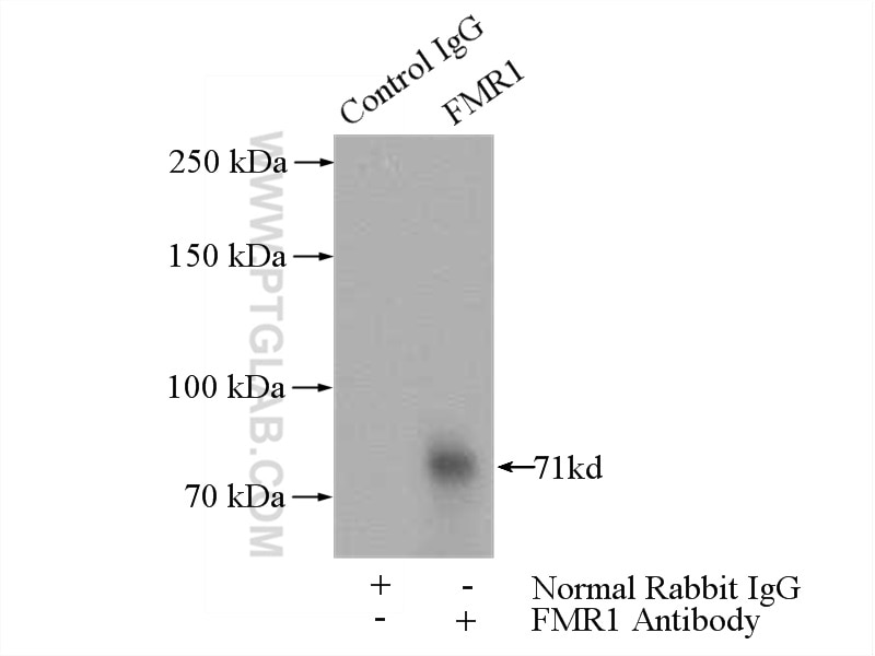
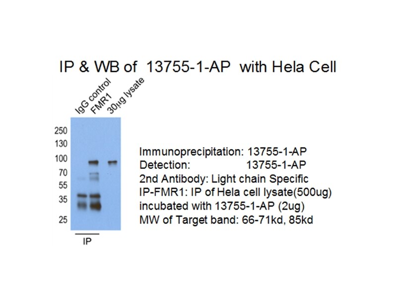
| Résultats positifs en IP | cellules HeLa |
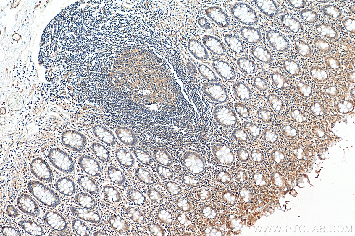
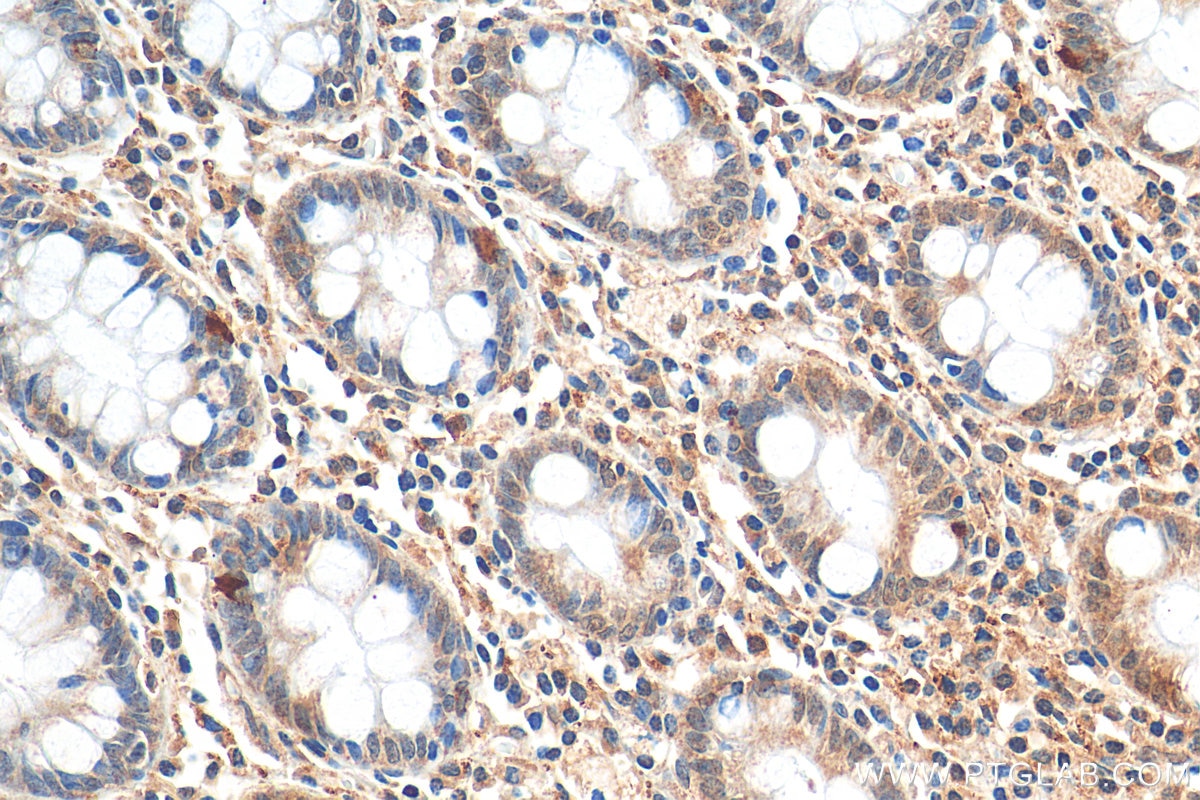
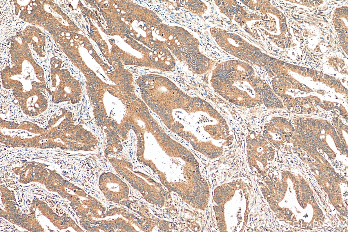
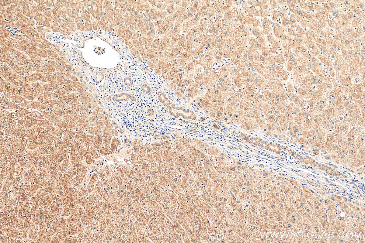
| Résultats positifs en IHC | tissu de côlon humain, tissu de cancer du côlon humain, tissu de gliome humain, tissu hépatique humain il est suggéré de démasquer l'antigène avec un tampon de TE buffer pH 9.0; (*) À défaut, 'le démasquage de l'antigène peut être 'effectué avec un tampon citrate pH 6,0. |
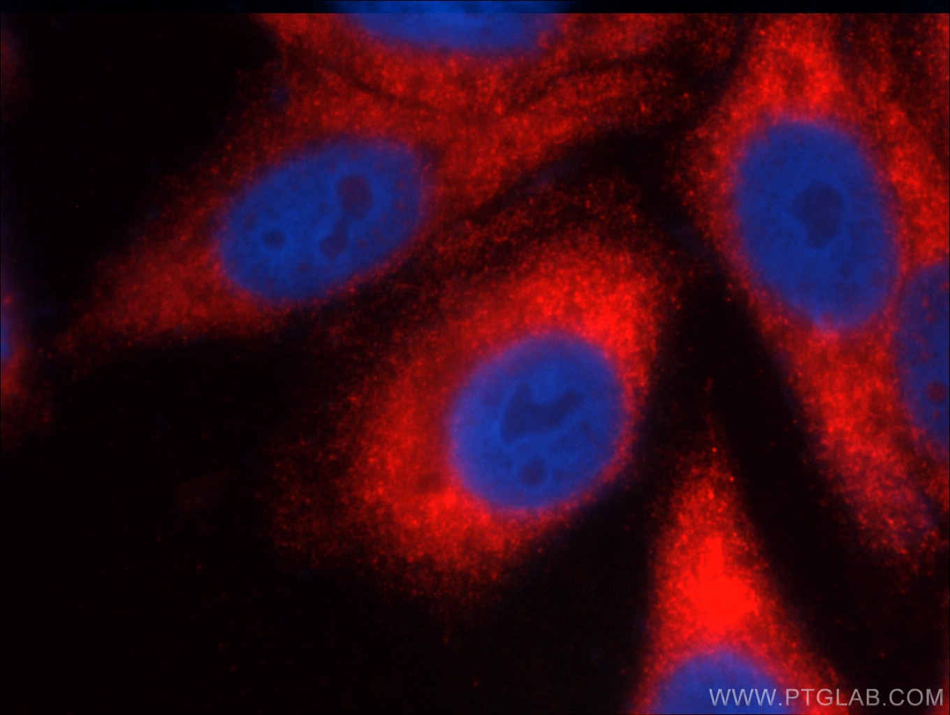
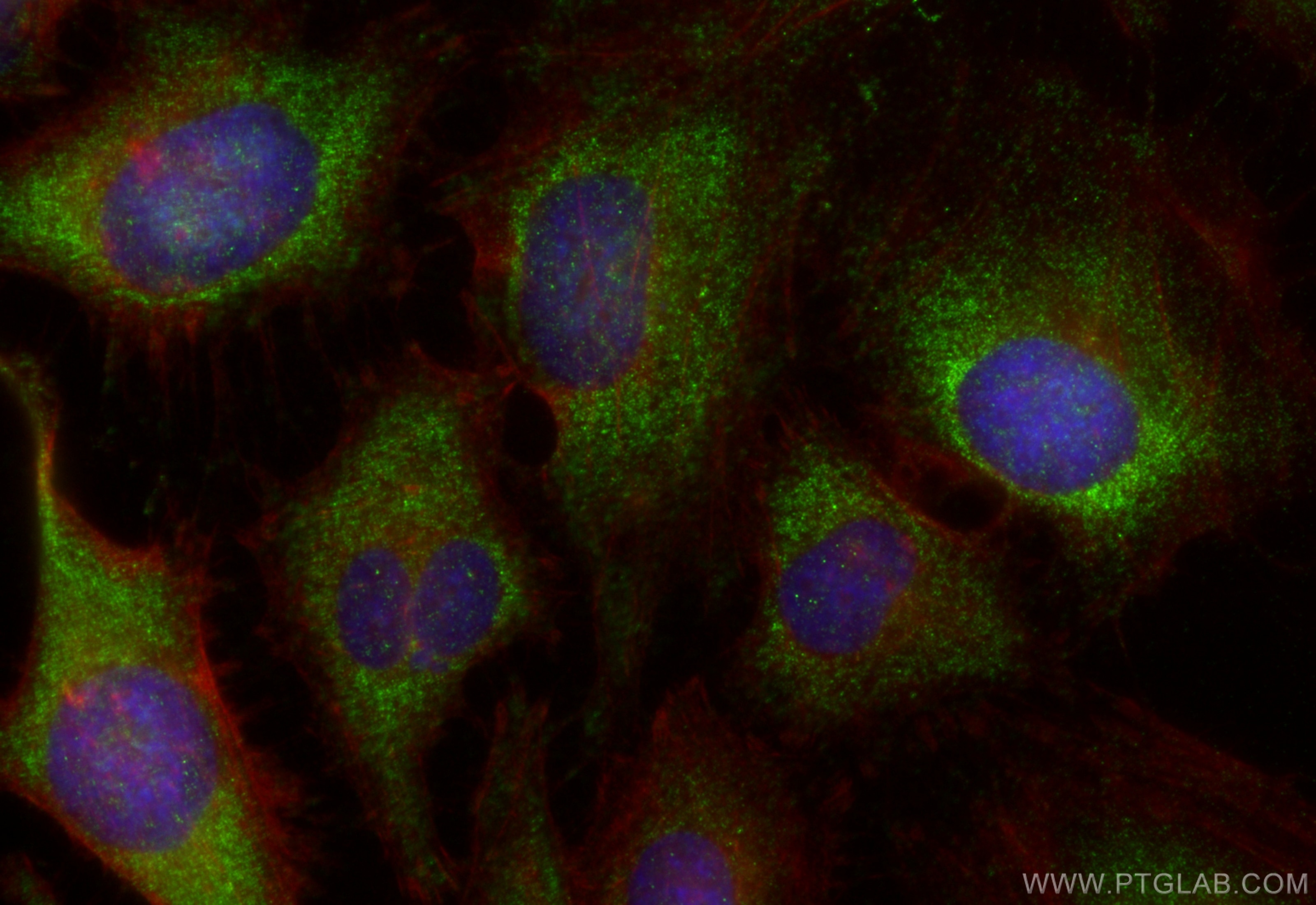
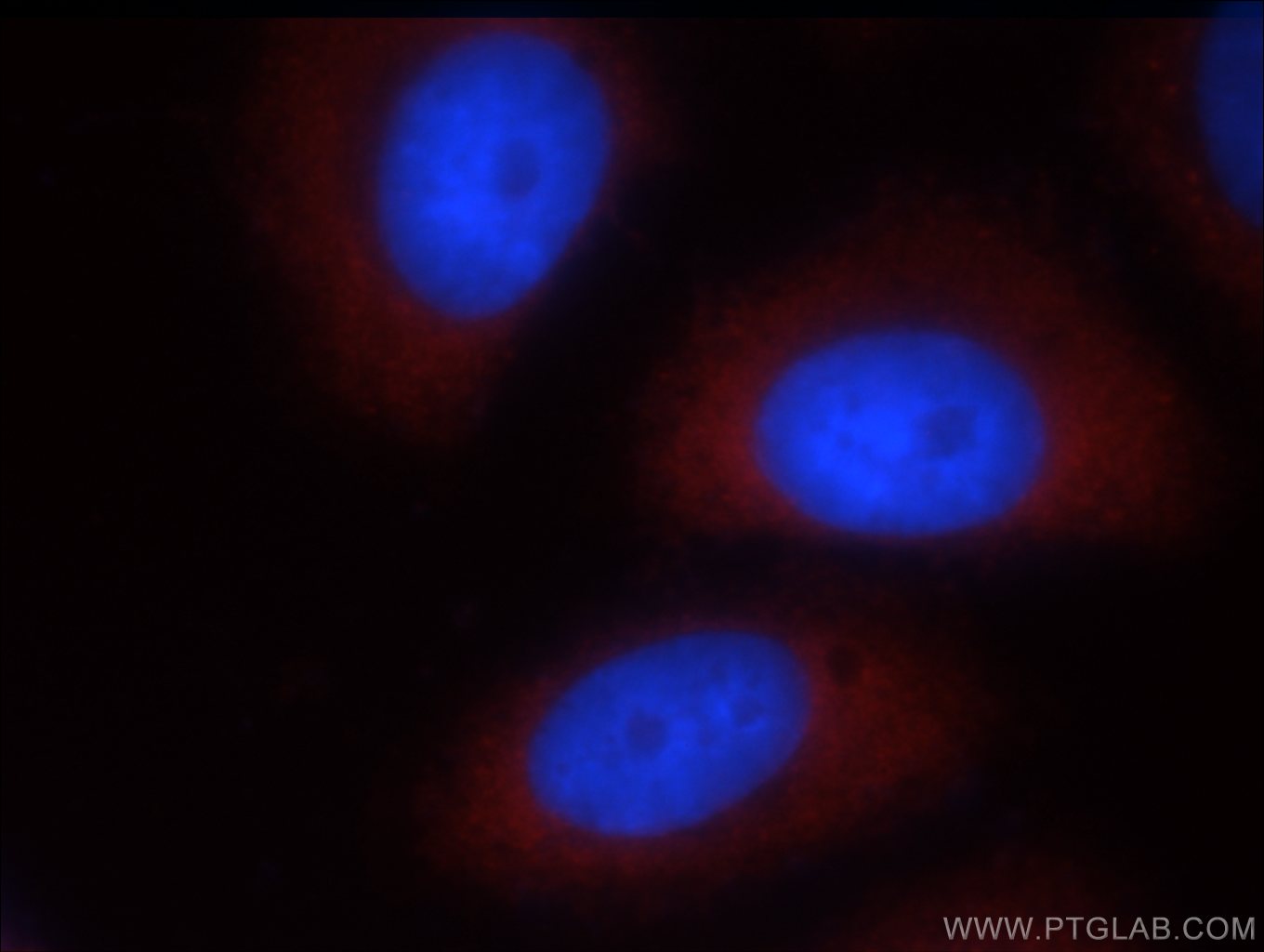
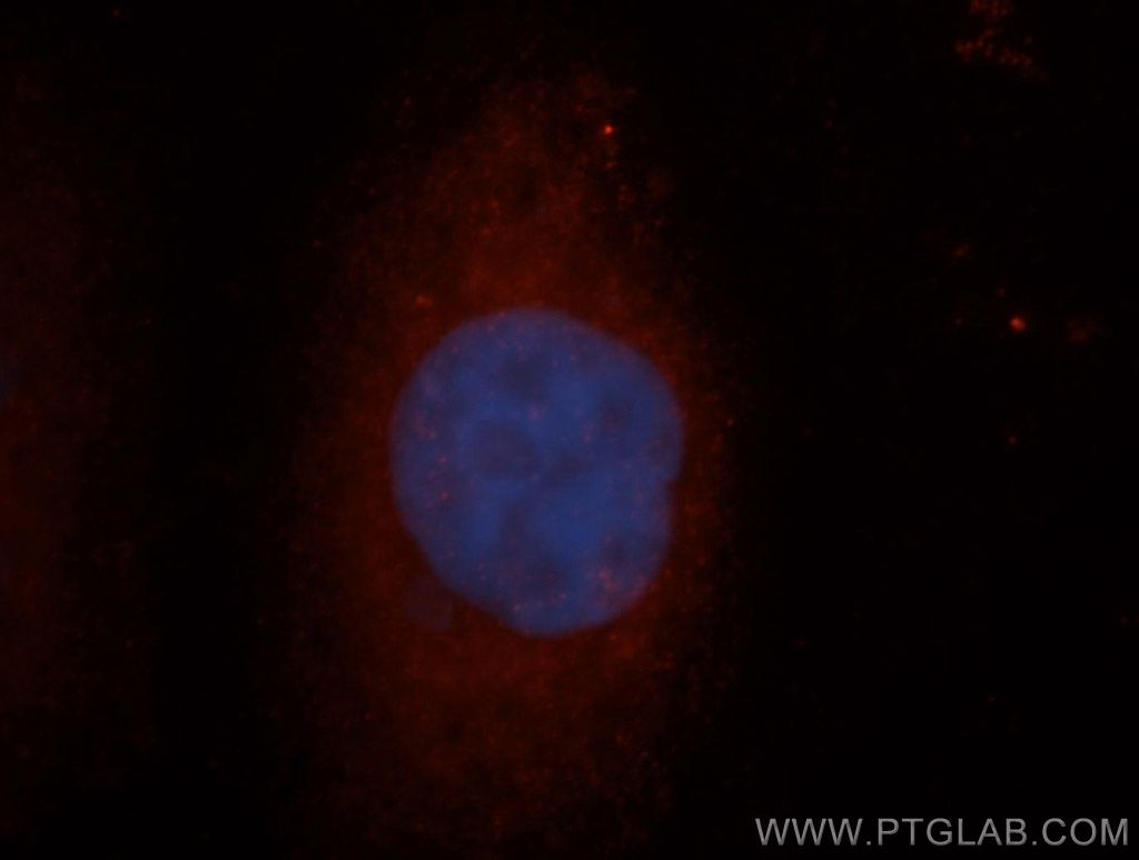
| Résultats positifs en IF/ICC | cellules HeLa, cellules HepG2 |
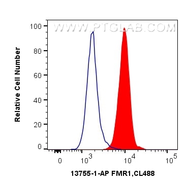
| Résultats positifs en FC (Intra) | cellules Jurkat, |
Dilution recommandée
| Application | Dilution |
|---|---|
| Western Blot (WB) | WB : 1:500-1:3000 |
| Immunoprécipitation (IP) | IP : 0.5-4.0 ug for 1.0-3.0 mg of total protein lysate |
| Immunohistochimie (IHC) | IHC : 1:200-1:800 |
| Immunofluorescence (IF)/ICC | IF/ICC : 1:50-1:500 |
| Flow Cytometry (FC) (INTRA) | FC (INTRA) : 0.40 ug per 10^6 cells in a 100 µl suspension |
| It is recommended that this reagent should be titrated in each testing system to obtain optimal results. | |
| Sample-dependent, check data in validation data gallery | |
Applications publiées
| KD/KO | See 2 publications below |
| WB | See 12 publications below |
| IHC | See 3 publications below |
| IF | See 2 publications below |
| IP | See 1 publications below |
Informations sur le produit
13755-1-AP cible FMR1 dans les applications de WB, IHC, IF/ICC, FC (Intra), IP, ELISA et montre une réactivité avec des échantillons Humain, souris
| Réactivité | Humain, souris |
| Réactivité citée | Humain, souris |
| Hôte / Isotype | Lapin / IgG |
| Clonalité | Polyclonal |
| Type | Anticorps |
| Immunogène | FMR1 Protéine recombinante Ag4697 |
| Nom complet | fragile X mental retardation 1 |
| Masse moléculaire calculée | 632 aa, 71 kDa |
| Poids moléculaire observé | 59-72 kDa |
| Numéro d’acquisition GenBank | BC038998 |
| Symbole du gène | FMR1 |
| Identification du gène (NCBI) | 2332 |
| Conjugaison | Non conjugué |
| Forme | Liquide |
| Méthode de purification | Purification par affinité contre l'antigène |
| Tampon de stockage | PBS with 0.02% sodium azide and 50% glycerol |
| Conditions de stockage | Stocker à -20°C. Stable pendant un an après l'expédition. L'aliquotage n'est pas nécessaire pour le stockage à -20oC Les 20ul contiennent 0,1% de BSA. |
Informations générales
The selective RNA-binding protein FMRP forms a messenger ribonucleoprotein complex that associates with polyribosomes, implicating in regulation of translation. FMR1 is a component of the CYFIP1-EIF4E-FMR1 complex which binds to and reperss the mRNA. It also has a role in the transport of mRNA from the nucleus to the cytoplasm. FMR1 exists several isforms and the molecular weight of FMR1 is about 59-72 kDa.
Protocole
| Product Specific Protocols | |
|---|---|
| WB protocol for FMR1 antibody 13755-1-AP | Download protocol |
| IHC protocol for FMR1 antibody 13755-1-AP | Download protocol |
| IF protocol for FMR1 antibody 13755-1-AP | Download protocol |
| IP protocol for FMR1 antibody 13755-1-AP | Download protocol |
| Standard Protocols | |
|---|---|
| Click here to view our Standard Protocols |
Publications
| Species | Application | Title |
|---|---|---|
Cell Competing Protein-RNA Interaction Networks Control Multiphase Intracellular Organization. | ||
Nat Commun Stalled translation by mitochondrial stress upregulates a CNOT4-ZNF598 ribosomal quality control pathway important for tissue homeostasis | ||
Cell Rep hnRNPA2B1 represses the disassembly of arsenite-induced stress granules and is essential for male fertility | ||
Int Immunopharmacol MNSFβ promotes LPS-induced TNFα expression by increasing the localization of RC3H1 to stress granules, and the interfering peptide HEPN2 reduces TNFα production by disrupting the MNSFβ-RC3H1 interaction in macrophages | ||
Cancer Cell Int MiR-323a-3p acts as a tumor suppressor by suppressing FMR1 and predicts better esophageal squamous cell carcinoma outcome. | ||
Biology (Basel) In Situ Peroxidase Labeling Followed by Mass-Spectrometry Reveals TIA1 Interactome. |
Avis
The reviews below have been submitted by verified Proteintech customers who received an incentive for providing their feedback.
FH Parisa (Verified Customer) (09-26-2023) | We used for WB, nor high specificity as we could see different bands (they could be different isoforms of the protein). But it was sensitive and we could see the band pulled down in IP for our protein of interest.
|
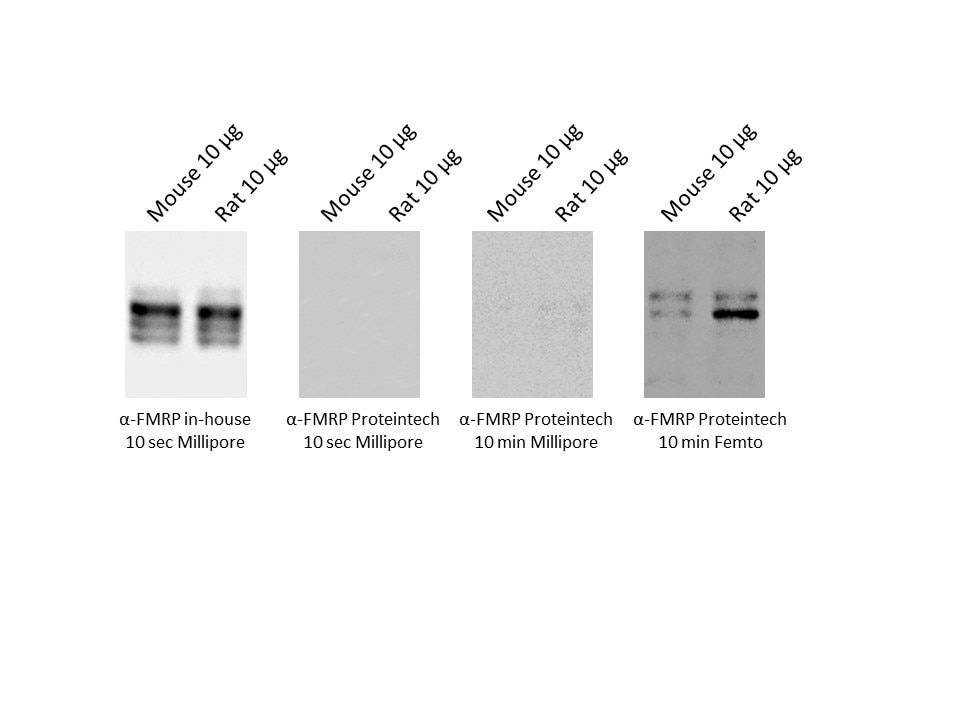
FH Marta (Verified Customer) (02-24-2020) | not specific enough in WB. Not tested in IPworst than the antibody that we have available in the antibody. Need a long exposure with a high sensible ECL.
 |
