- Phare
- Validé par KD/KO
Anticorps Polyclonal de lapin anti-GTPBP4
GTPBP4 Polyclonal Antibody for WB, IF/ICC, IP, ELISA
Hôte / Isotype
Lapin / IgG
Réactivité testée
Humain, souris et plus (2)
Applications
WB, IHC, IF/ICC, IP, ELISA
Conjugaison
Non conjugué
N° de cat : 13897-1-AP
Synonymes
Galerie de données de validation
Applications testées
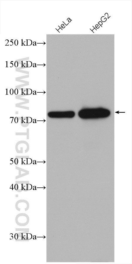
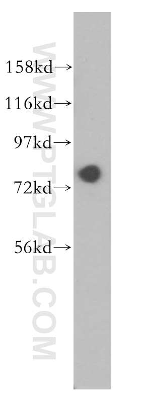
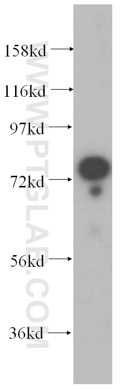
| Résultats positifs en WB | cellules HeLa, cellules HepG2, tissu rénal humain, tissu testiculaire de souris |
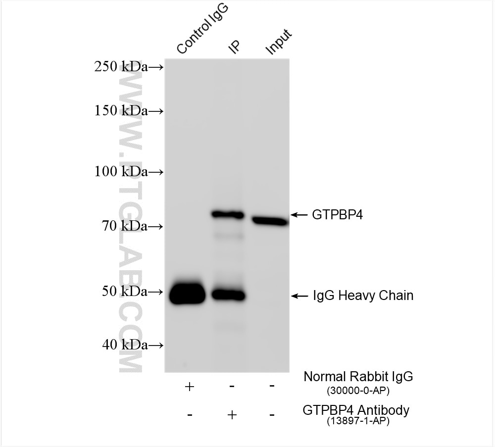
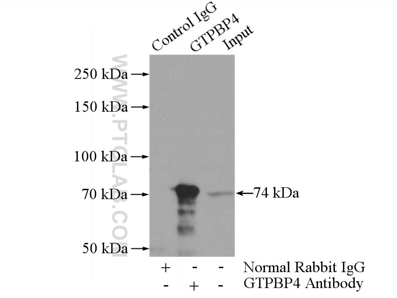
| Résultats positifs en IP | cellules HeLa, tissu testiculaire de souris |
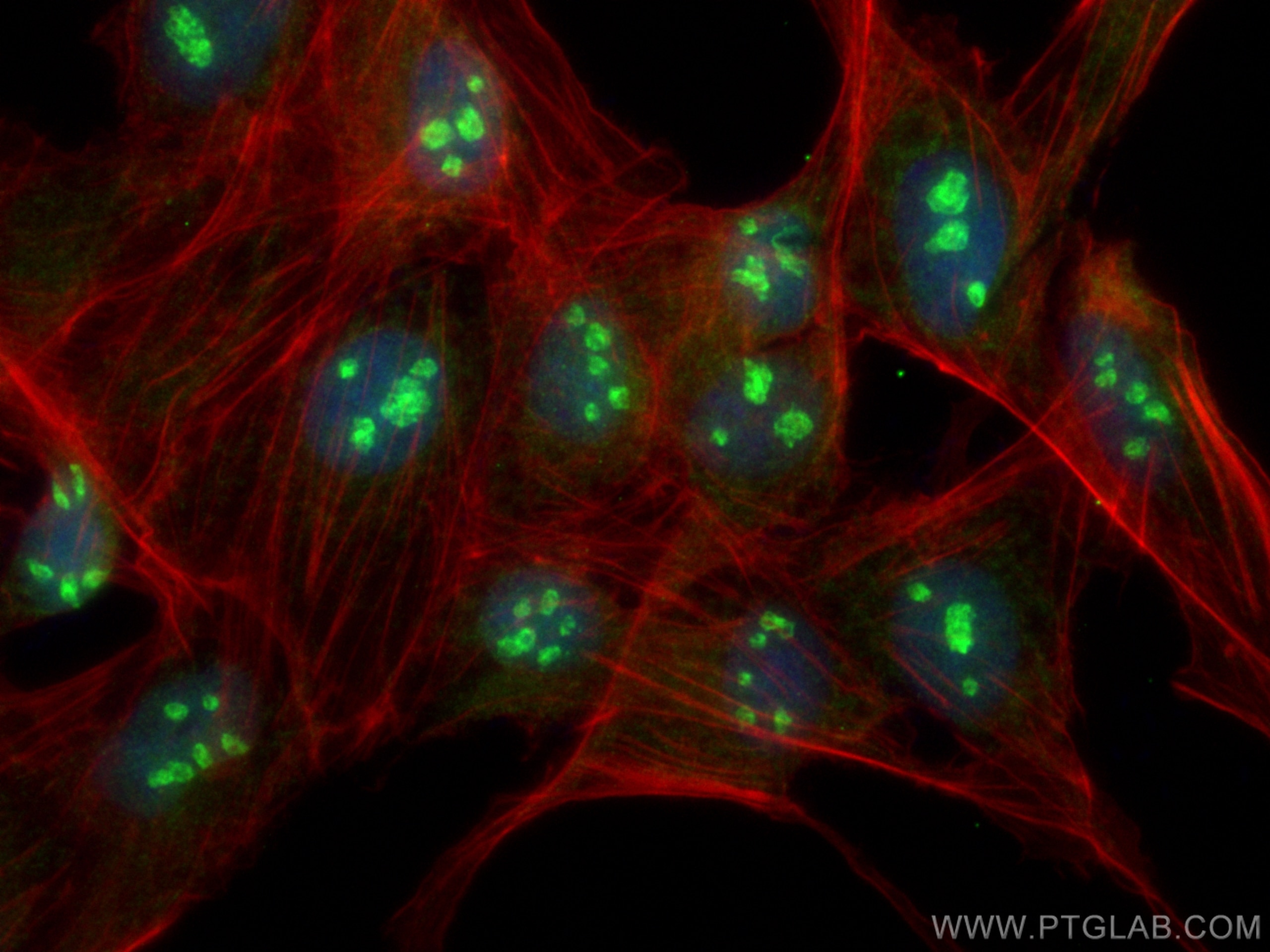
| Résultats positifs en IF/ICC | cellules MDCK, |
Dilution recommandée
| Application | Dilution |
|---|---|
| Western Blot (WB) | WB : 1:2000-1:8000 |
| Immunoprécipitation (IP) | IP : 0.5-4.0 ug for 1.0-3.0 mg of total protein lysate |
| Immunofluorescence (IF)/ICC | IF/ICC : 1:200-1:800 |
| It is recommended that this reagent should be titrated in each testing system to obtain optimal results. | |
| Sample-dependent, check data in validation data gallery | |
Applications publiées
| KD/KO | See 3 publications below |
| WB | See 6 publications below |
| IHC | See 1 publications below |
| IF | See 2 publications below |
Informations sur le produit
13897-1-AP cible GTPBP4 dans les applications de WB, IHC, IF/ICC, IP, ELISA et montre une réactivité avec des échantillons Humain, souris
| Réactivité | Humain, souris |
| Réactivité citée | Chèvre, Drosophile, Humain, souris |
| Hôte / Isotype | Lapin / IgG |
| Clonalité | Polyclonal |
| Type | Anticorps |
| Immunogène | GTPBP4 Protéine recombinante Ag4859 |
| Nom complet | GTP binding protein 4 |
| Masse moléculaire calculée | 634 aa, 74 kDa |
| Poids moléculaire observé | 74 kDa |
| Numéro d’acquisition GenBank | BC038975 |
| Symbole du gène | GTPBP4 |
| Identification du gène (NCBI) | 23560 |
| Conjugaison | Non conjugué |
| Forme | Liquide |
| Méthode de purification | Purification par affinité contre l'antigène |
| Tampon de stockage | PBS with 0.02% sodium azide and 50% glycerol |
| Conditions de stockage | Stocker à -20°C. Stable pendant un an après l'expédition. L'aliquotage n'est pas nécessaire pour le stockage à -20oC Les 20ul contiennent 0,1% de BSA. |
Informations générales
GTP-binding proteins are GTPases and function as molecular switches that can flip between two states: active, when GTP is bound, and inactive, when GDP is bound. 'Active' in this context usually means that the molecule acts as a signal to trigger other events in the cell. When an extracellular ligand binds to a G-protein-linked receptor, the receptor changes its conformation and switches on the trimeric G proteins that associate with it by causing them to eject their GDP and replace it with GTP. The switch is turned off when the G protein hydrolyzes its own bound GTP, converting it back to GDP. But before that occurs, the active protein has an opportunity to diffuse away from the receptor and deliver its message for a prolonged period to its downstream target.
Protocole
| Product Specific Protocols | |
|---|---|
| WB protocol for GTPBP4 antibody 13897-1-AP | Download protocol |
| IF protocol for GTPBP4 antibody 13897-1-AP | Download protocol |
| IP protocol for GTPBP4 antibody 13897-1-AP | Download protocol |
| Standard Protocols | |
|---|---|
| Click here to view our Standard Protocols |
Publications
| Species | Application | Title |
|---|---|---|
Genes Dev Uncoupling of GTP hydrolysis from eIF6 release on the ribosome causes Shwachman-Diamond syndrome. | ||
Proc Natl Acad Sci U S A A genome-scale protein interaction profile of Drosophila p53 uncovers additional nodes of the human p53 network.
| ||
Biochem Biophys Res Commun Up-regulation of GTPBP4 in colorectal carcinoma is responsible for tumor metastasis.
| ||
Biomed Res Int Determining the Clinical Value and Critical Pathway of GTPBP4 in Lung Adenocarcinoma Using a Bioinformatics Strategy: A Study Based on Datasets from The Cancer Genome Atlas.
| ||
Cell Rep UTP18-mediated p21 mRNA instability drives adenoma-carcinoma progression in colorectal cancer | ||
Nucleic Acids Res Distinct interactomes of ADAR1 nuclear and cytoplasmic protein isoforms and their responses to interferon induction |
Avis
The reviews below have been submitted by verified Proteintech customers who received an incentive for providing their feedback.
FH kaosheng (Verified Customer) (11-11-2019) | This antibody detects a very clear and specific band in polysome profiling fractions of human TF1 cells.
|
