- Phare
- Validé par KD/KO
Anticorps Polyclonal de lapin anti-HIF-1 alpha
HIF-1 alpha Polyclonal Antibody for WB, IHC, IF/ICC, IP, ELISA
Hôte / Isotype
Lapin / IgG
Réactivité testée
Humain et plus (3)
Applications
WB, IHC, IF/ICC, IP, CoIP, ChIP, RIP, ELISA
Conjugaison
Non conjugué
N° de cat : 20960-1-AP
Synonymes
Galerie de données de validation
 at dilution of 1:6000 incubated at room temperature for 1.5 hours.")
 at dilution of 1:6000 incubated at room temperature for 1.5 hours.")
 with sh-Control and sh-HIF-1 alpha transfected HeLa cells. Sample 1: non-treated sh-Control transfected HeLa cells, Sample 2: Cobalt Chloride treated sh-Control transfected HeLa cells, Sample 3: Cobalt Chloride treated sh-HIF-1 alpha transfected HeLa cells.")
 at dilution of 1:4000 incubated at room temperature for 1.5 hours.")
 with HeLa cells lysate 4000ug.")
 at dilution of 1:200 (under 40x lens). Heat mediated antigen retrieval with Tris-EDTA buffer (pH 9.0).")
 at dilution of 1:200 (under 10x lens). Heat mediated antigen retrieval with Tris-EDTA buffer (pH 9.0).")
 at dilution of 1:100 (under 10x lens).")
 at dilution of 1:100 (under 40x lens).")
 at dilution of 1:50 (under 10x lens).")
 at dilution of 1:50 (under 40x lens).")
 at dilution of 1:50 (under 10x lens).")
 at dilution of 1:50 (under 40x lens).")
 fixed Cobalt Chloride treated HeLa cells using HIF-1 alpha antibody (20960-1-AP) at dilution of 1:400 and CoraLite®488-Conjugated AffiniPure Goat Anti-Rabbit IgG(H+L), CL594-phalloidin (red).")
 fixed Cobalt Chloride treated HepG2 cells using HIF1a antibody (20960-1-AP) at dilution of 1:1800 and CoraLite®488-Conjugated AffiniPure Goat Anti-Rabbit IgG(H+L).")
 fixed Cobalt Chloride treated HepG2 cells using HIF1a antibody (20960-1-AP) at dilution of 1:500 and CoraLite®488-Conjugated AffiniPure Goat Anti-Rabbit IgG(H+L).")
 fixed Cobalt Chloride treated HeLa cells using HIF-1 alpha antibody (20960-1-AP) at dilution of 1:400 and CoraLite®488-Conjugated AffiniPure Goat Anti-Rabbit IgG(H+L), CL594-phalloidin (red).")
Applications testées
| Résultats positifs en WB | cellules HeLa traitées au chlorure de cobalt, cellules HeLa |
| Résultats positifs en IP | cellules HeLa, |
| Résultats positifs en IHC | tissu de cancer de la thyroïde humain, tissu cardiaque humain, tissu rénal humain il est suggéré de démasquer l'antigène avec un tampon de TE buffer pH 9.0; (*) À défaut, 'le démasquage de l'antigène peut être 'effectué avec un tampon citrate pH 6,0. |
| Résultats positifs en IF/ICC | cellules HeLa traitées au chlorure de cobalt, cellules HepG2 traitées au chlorure de cobalt |
Dilution recommandée
| Application | Dilution |
|---|---|
| Western Blot (WB) | WB : 1:2000-1:12000 |
| Immunoprécipitation (IP) | IP : 0.5-4.0 ug for 1.0-3.0 mg of total protein lysate |
| Immunohistochimie (IHC) | IHC : 1:50-1:500 |
| Immunofluorescence (IF)/ICC | IF/ICC : 1:200-1:800 |
| It is recommended that this reagent should be titrated in each testing system to obtain optimal results. | |
| Sample-dependent, check data in validation data gallery | |
Informations sur le produit
20960-1-AP cible HIF-1 alpha dans les applications de WB, IHC, IF/ICC, IP, CoIP, ChIP, RIP, ELISA et montre une réactivité avec des échantillons Humain
| Réactivité | Humain |
| Réactivité citée | Chèvre, Humain, porc, poulet |
| Hôte / Isotype | Lapin / IgG |
| Clonalité | Polyclonal |
| Type | Anticorps |
| Immunogène | HIF-1 alpha Protéine recombinante Ag15198 |
| Nom complet | hypoxia inducible factor 1, alpha subunit (basic helix-loop-helix transcription factor) |
| Masse moléculaire calculée | 826 aa, 93 kDa |
| Poids moléculaire observé | 120 kDa |
| Numéro d’acquisition GenBank | BC012527 |
| Symbole du gène | HIF1A |
| Identification du gène (NCBI) | 3091 |
| Conjugaison | Non conjugué |
| Forme | Liquide |
| Méthode de purification | Purification par affinité contre l'antigène |
| Tampon de stockage | PBS with 0.02% sodium azide and 50% glycerol |
| Conditions de stockage | Stocker à -20°C. Stable pendant un an après l'expédition. L'aliquotage n'est pas nécessaire pour le stockage à -20oC Les 20ul contiennent 0,1% de BSA. |
Informations générales
HIF1a, the major regulator of the cellular responses to hypoxia, consists of an oxygen-sensitive subunit, HIF1 alpha (HIF1A), and an oxygen-insensitive subunit, HIF1 beta (arylhydrocarbon receptor nuclear transporter [ARNT]). Under normal oxygen conditions, HIF1a is continuously produced and destroyed, in a process involving hydroxylation, interaction with von Hippel-Lindau (VHL) protein, polyubiquitylation and subsequent proteasomal degradation. Under hypoxic conditions, hydroxylation is impaired and HIF1a is stabilized. HIF1a localizes in cytoplasm in normoxia, but it can translocate into nuclear in response to hypoxia. The calculated molecular weight of HIF1a is 93 kDa, but the modified protein HIF1a is about 110-120kDa (PMID: 11698256, .PMID: 7539918). .
Protocole
| Product Specific Protocols | |
|---|---|
| WB protocol for HIF-1 alpha antibody 20960-1-AP | Download protocol |
| IHC protocol for HIF-1 alpha antibody 20960-1-AP | Download protocol |
| IF protocol for HIF-1 alpha antibody 20960-1-AP | Download protocol |
| IP protocol for HIF-1 alpha antibody 20960-1-AP | Download protocol |
| Standard Protocols | |
|---|---|
| Click here to view our Standard Protocols |
Publications
| Species | Application | Title |
|---|---|---|
Gastroenterology Pancreatic acinar cells-derived sphingosine-1-phosphate contributes to fibrosis of chronic pancreatitis via inducing autophagy and activation of pancreatic stellate cells | ||
Cancer Cell Cancer cell autophagy, reprogrammed macrophages, and remodeled vasculature in glioblastoma triggers tumor immunity | ||
Nat Cell Biol A single-cell transcriptomic landscape of the lungs of patients with COVID-19. | ||
ACS Nano Augmenting Intracellular Cargo Delivery of Extracellular Vesicles in Hypoxic Tissues through Inhibiting Hypoxia-Induced Endocytic Recycling | ||
Mol Cell The mitochondrial DNAJC co-chaperone TCAIM reduces α-ketoglutarate dehydrogenase protein levels to regulate metabolism |
Avis
The reviews below have been submitted by verified Proteintech customers who received an incentive for providing their feedback.
FH Vignesh (Verified Customer) (09-15-2025) | The results are excellent.
|
FH Carlo (Verified Customer) (01-18-2023) | The quality of the band in western blot is well defined
|
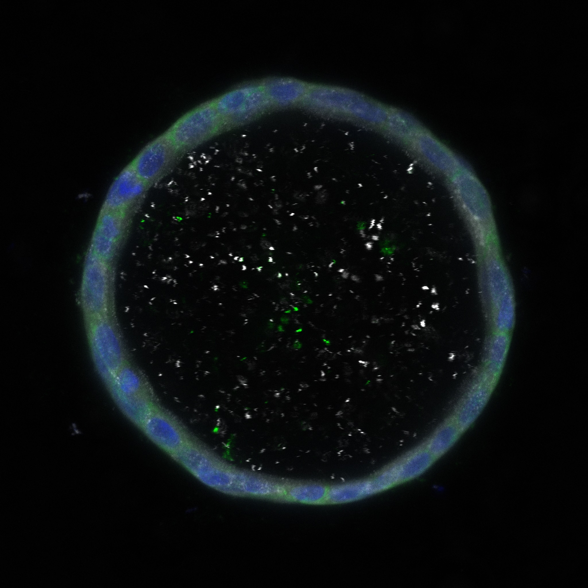
FH Davide (Verified Customer) (09-20-2022) | The image uploaded refers to a hypoxic condition O2<1%. The antibody signal was aspecific and generated aspecific binding in the middle of the organoid.
 |
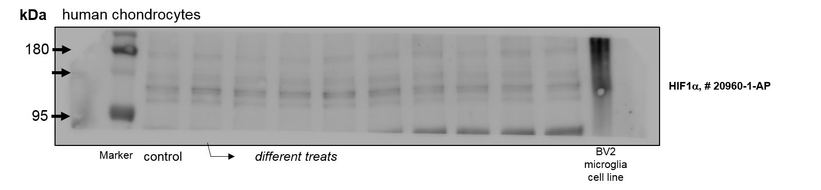
FH Christian (Verified Customer) (01-07-2022) | Unfortunately much background an many unspecific bands in lysates of human chondrocytes and BV2 microclia cells (less unspecific bands in rat chondrocytes).
 |
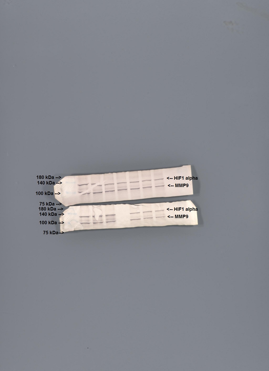
FH Diane (Verified Customer) (02-17-2021) | Separated on a 7.5% SDS PAGE gel, transferred to nitrocellulose membrane. Block with 5% dry milk/PBST at room temperature for one hour. 1:1000 dilution of the primary antibody in blocking solution. Incubated overnight at 4 degrees C on a rocker platform. Secondary antibody 1:1000 secondary (anti rabbit HRP) incubation for two hours at room temperature. Visualization with the Opti-4CN chromogenic substrate kit (BioRad).
 |
FH Chun (Verified Customer) (12-05-2019) | A good antibody
|

