- Phare
- Validé par KD/KO
Anticorps Polyclonal de lapin anti-HSP70
HSP70 Polyclonal Antibody for WB, IHC, IF/ICC, FC (Intra), IP, ELISA
Hôte / Isotype
Lapin / IgG
Réactivité testée
Humain, rat, souris et plus (5)
Applications
WB, IHC, IF/ICC, FC (Intra), IP, CoIP, ELISA
Conjugaison
Non conjugué
N° de cat : 10995-1-AP
Synonymes
Galerie de données de validation
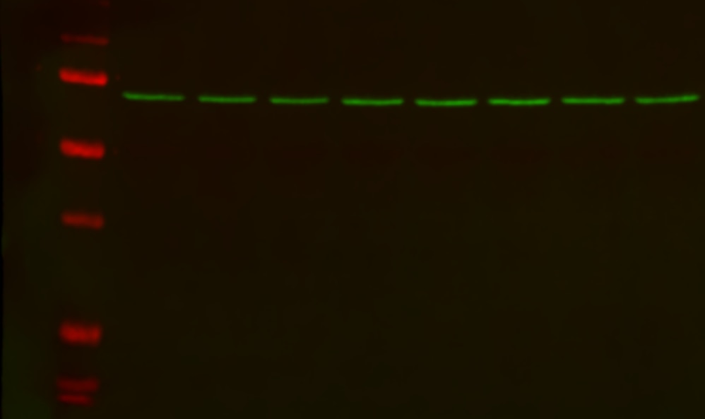
 at dilution of 1:10000 incubated at room temperature for 1.5 hours.")
 at dilution of 1:10000 incubated at room temperature for 1.5 hours.")
 at dilution of 1:10000 incubated at room temperature for 1.5 hours.")
 with mouse brain tissue lysate 3600ug.")
 at dilution of 1:200 (under 10x lens). Heat mediated antigen retrieval with Tris-EDTA buffer (pH 9.0).")
 at dilution of 1:2000 (under 20x lens). Heat mediated antigen retrieval with Tris-EDTA buffer (pH 9.0).")
 at dilution of 1:2000 (under 20x lens). Heat mediated antigen retrieval with Tris-EDTA buffer (pH 9.0).")
 at dilution of 1:200 (under 40x lens).")
 at dilution of 1:200 (under 10x lens). Heat mediated antigen retrieval with Tris-EDTA buffer (pH 9.0).")
 at dilution of 1:200 (under 10x lens). Heat mediated antigen retrieval with Tris-EDTA buffer (pH 9.0).")
 fixed HeLa cells using HSP70 antibody (10995-1-AP) at dilution of 1:400 and CoraLite®488-Conjugated AffiniPure Goat Anti-Rabbit IgG(H+L).")
. Blue pseudocolor = DAPI (fluorescent DNA dye).")
 and CoraLite®488-Conjugated Goat Anti-Rabbit IgG(H+L) (SA00013-2)(red), or 0.25 ug rabbit IgG isotype control (blue). Cells were fixed with 4% PFA and permeabilized with Flow Cytometry Perm Buffer (PF00011-C).")
Applications testées
| Résultats positifs en WB | cellules A431, cellules A549, cellules HEK-293, cellules HeLa, cellules Jurkat, cerveau de souris, rein de rat, rein de souris |
| Résultats positifs en IP | tissu cérébral de souris |
| Résultats positifs en IHC | tissu de cancer du sein humain, tissu cutané de souris, tissu de cancer du foie humain il est suggéré de démasquer l'antigène avec un tampon de TE buffer pH 9.0; (*) À défaut, 'le démasquage de l'antigène peut être 'effectué avec un tampon citrate pH 6,0. |
| Résultats positifs en IF/ICC | cellules HeLa, cellules HepG2 |
| Résultats positifs en FC (Intra) | cellules HeLa, |
Dilution recommandée
| Application | Dilution |
|---|---|
| Western Blot (WB) | WB : 1:5000-1:50000 |
| Immunoprécipitation (IP) | IP : 0.5-4.0 ug for 1.0-3.0 mg of total protein lysate |
| Immunohistochimie (IHC) | IHC : 1:200-1:2000 |
| Immunofluorescence (IF)/ICC | IF/ICC : 1:200-1:800 |
| Flow Cytometry (FC) (INTRA) | FC (INTRA) : 0.25 ug per 10^6 cells in a 100 µl suspension |
| It is recommended that this reagent should be titrated in each testing system to obtain optimal results. | |
| Sample-dependent, check data in validation data gallery | |
Applications publiées
| KD/KO | See 9 publications below |
| WB | See 302 publications below |
| IHC | See 13 publications below |
| IF | See 26 publications below |
| IP | See 1 publications below |
| ELISA | See 1 publications below |
| CoIP | See 5 publications below |
Informations sur le produit
10995-1-AP cible HSP70 dans les applications de WB, IHC, IF/ICC, FC (Intra), IP, CoIP, ELISA et montre une réactivité avec des échantillons Humain, rat, souris
| Réactivité | Humain, rat, souris |
| Réactivité citée | rat, Chèvre, Humain, Lapin, poisson-zèbre, poulet, souris, oyster |
| Hôte / Isotype | Lapin / IgG |
| Clonalité | Polyclonal |
| Type | Anticorps |
| Immunogène | HSP70 Protéine recombinante Ag1446 |
| Nom complet | heat shock 70kDa protein 1A |
| Masse moléculaire calculée | 70 kDa |
| Poids moléculaire observé | 66-70 kDa |
| Numéro d’acquisition GenBank | BC009322 |
| Symbole du gène | HSP70 |
| Identification du gène (NCBI) | 3303 |
| Conjugaison | Non conjugué |
| Forme | Liquide |
| Méthode de purification | Purification par affinité contre l'antigène |
| Tampon de stockage | PBS with 0.02% sodium azide and 50% glycerol |
| Conditions de stockage | Stocker à -20°C. Stable pendant un an après l'expédition. L'aliquotage n'est pas nécessaire pour le stockage à -20oC Les 20ul contiennent 0,1% de BSA. |
Informations générales
-
What is Hsp70/HSP1A?
HSP1A is a member of the Hsp70 (heat shock protein 70) proteins that act as molecular chaperones ensuring correct protein folding and preventing protein aggregation. Hsp70 protein production is greatly induced by various stress stimuli, including high temperature and toxins. Its expression is often elevated in various cancers.
-
FAQs for Hsp70
a. I cannot detect Hsp70 by western blotting
HSP70s are typically expressed at low levels under normal physiological conditions but are dramatically up-regulated in response to cellular stress. Try to always include cell lysate from cells subjected to stress conditions as a positive control.
b. What loading control can I use for cellular stress experiments with Hsp70?
Choosing a loading control antibody is an important step in western blotting experimental setup. We highly recommend using more than one loading control while developing new cellular stress assays to ensure that a given treatment does not alter expression of house-keeping genes. Hsp70 has a molecular size of 70 kDa, so we recommend using GAPDH (36 kDa), actin (42 kDa), or tubulin (50-55 kDa). More information on our control antibodies can be found here: https://www.ptglab.com/news/blog/loading-control-antibodies-for-western-blotting/.
Protocole
| Product Specific Protocols | |
|---|---|
| WB protocol for HSP70 antibody 10995-1-AP | Download protocol |
| IHC protocol for HSP70 antibody 10995-1-AP | Download protocol |
| IF protocol for HSP70 antibody 10995-1-AP | Download protocol |
| IP protocol for HSP70 antibody 10995-1-AP | Download protocol |
| Standard Protocols | |
|---|---|
| Click here to view our Standard Protocols |
Publications
| Species | Application | Title |
|---|---|---|
Gastroenterology PTEN deficiency facilitates exosome secretion and metastasis in cholangiocarcinoma by impairing TFEB-mediated lysosome biogenesis | ||
Nat Cell Biol Caspase-2 is a condensate-mediated deubiquitinase in protein quality control | ||
Nat Cell Biol Heat stress activates YAP/TAZ to induce the heat shock transcriptome.
| ||
Mol Cancer CircEZH2/miR-133b/IGF2BP2 aggravates colorectal cancer progression via enhancing the stability of m6A-modified CREB1 mRNA. | ||
Adv Sci (Weinh) Reprogramming Lung Redox Homeostasis by NIR Driven Ultra-Small Pd Loaded Covalent Organic Framework Inhibits NF-κB Pathway for Acute Lung Injury Immunotherapy |
Avis
The reviews below have been submitted by verified Proteintech customers who received an incentive for providing their feedback.
FH PK (Verified Customer) (08-14-2024) |
 |
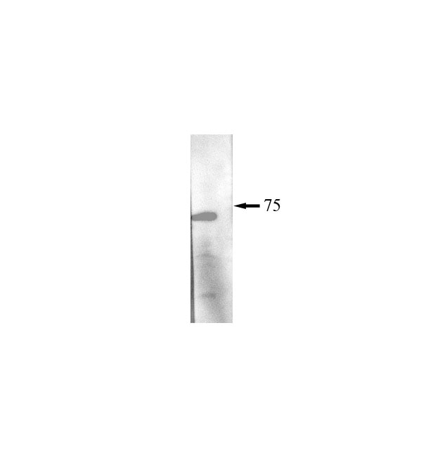
FH Scott (Verified Customer) (05-22-2024) | 20µg of protein was loaded and antibody was incubated overnight at 4oC following a total protein stain. The band appeared at the expected size. Precision plus protein standard ladder #1610373.
 |
FH Nin (Verified Customer) (01-28-2023) | A very good antibody to probe HSP70 by WB.
 |
FH Tsimafei (Verified Customer) (12-04-2022) | 20µg of the total protein were used. Incubation was done for 2h at room temperature
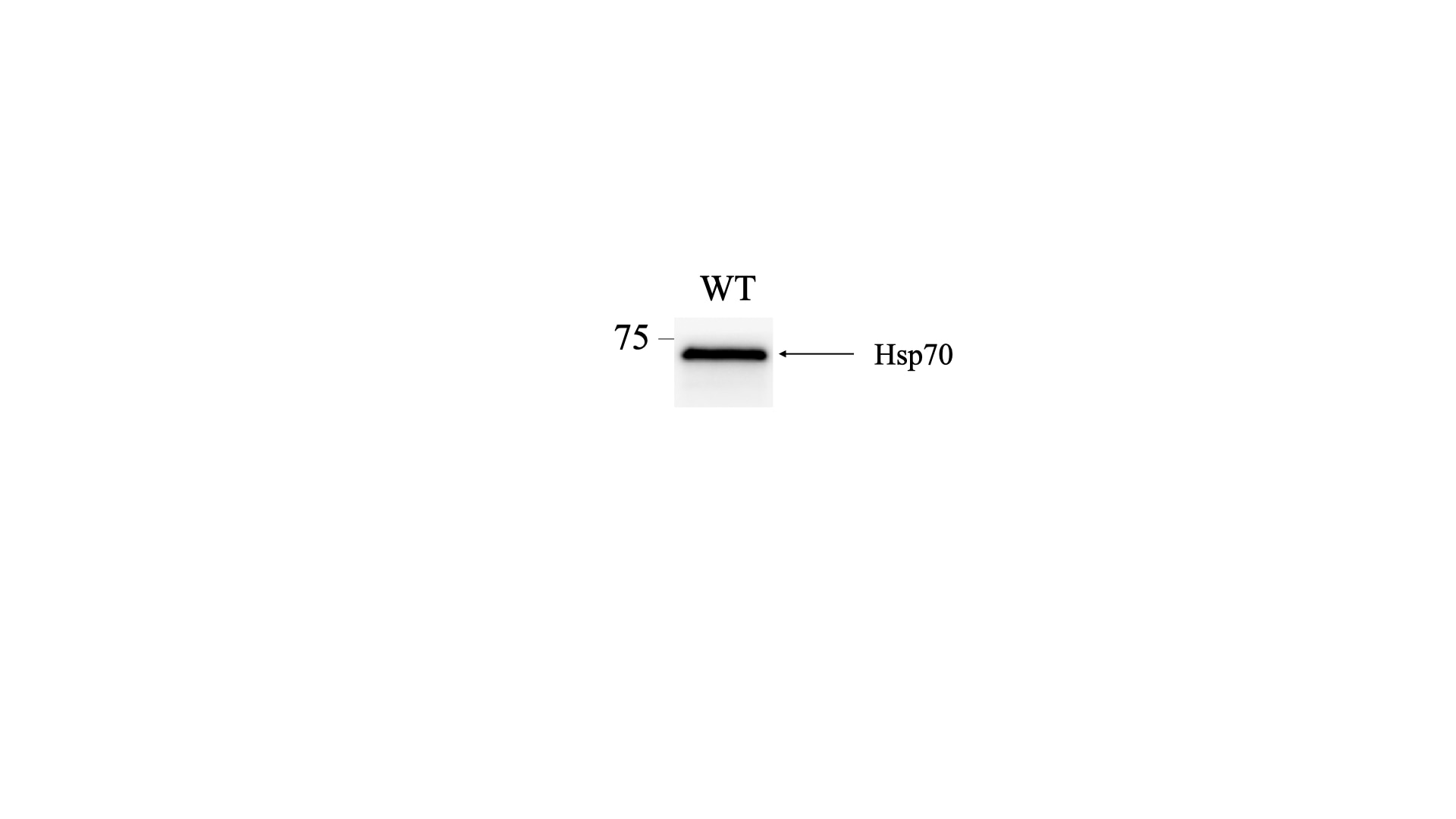 |
FH Guorong (Verified Customer) (03-22-2022) | Excellent performance with a specific band of expected size
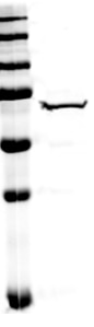 |
FH Luxi (Verified Customer) (11-23-2020) | This antibody works well in my tissue. It binds to the nuclear and cytosol Hsp70s.
|

