- Phare
- Validé par KD/KO
Anticorps Polyclonal de lapin anti-IDH3A
IDH3A Polyclonal Antibody for WB, IHC, IF/ICC, FC (Intra), IP, ELISA
Hôte / Isotype
Lapin / IgG
Réactivité testée
Humain, rat, souris
Applications
WB, IHC, IF/ICC, FC (Intra), IP, CoIP, ELISA
Conjugaison
Non conjugué
N° de cat : 15909-1-AP
Synonymes
Galerie de données de validation
 at dilution of 1:1200 incubated at room temperature for 1.5 hours.")
 at dilution of 1:10000 incubated at room temperature for 1.5 hours.")
 at dilution of 1:1000 incubated at room temperature for 1.5 hours.")
 at dilution of 1:1000 incubated at room temperature for 1.5 hours.")
 with HepG2 cells lysate 2400ug.")
 at dilution of 1:100 (under 40x lens).")
 at dilution of 1:100 (under 10x lens).")
 at dilution of 1:50 and Alexa Fluor 488-conjugated AffiniPure Goat Anti-Rabbit IgG(H+L).")
 fixed HepG2 cells using IDH3A antibody (15909-1-AP) at dilution of 1:400 and CoraLite®488-Conjugated AffiniPure Goat Anti-Rabbit IgG(H+L), CL594-phalloidin (red).")
 and CoraLite®488-Conjugated AffiniPure Goat Anti-Rabbit IgG(H+L) at dilution 1:1000 (red), or 0.4 ug Control Antibody. Cells were fixed with 4% PFA and permeabilized with Flow Cytometry Perm Buffer (PF00011-C).")
Applications testées
| Résultats positifs en WB | cellules HepG2, tissu cérébral de rat, tissu cérébral de souris, tissu de muscle squelettique humain |
| Résultats positifs en IP | cellules HepG2 |
| Résultats positifs en IHC | tissu cérébral humain il est suggéré de démasquer l'antigène avec un tampon de TE buffer pH 9.0; (*) À défaut, 'le démasquage de l'antigène peut être 'effectué avec un tampon citrate pH 6,0. |
| Résultats positifs en IF/ICC | cellules HepG2, |
| Résultats positifs en FC (Intra) | cellules HepG2, |
Dilution recommandée
| Application | Dilution |
|---|---|
| Western Blot (WB) | WB : 1:5000-1:50000 |
| Immunoprécipitation (IP) | IP : 0.5-4.0 ug for 1.0-3.0 mg of total protein lysate |
| Immunohistochimie (IHC) | IHC : 1:20-1:200 |
| Immunofluorescence (IF)/ICC | IF/ICC : 1:200-1:800 |
| Flow Cytometry (FC) (INTRA) | FC (INTRA) : 0.40 ug per 10^6 cells in a 100 µl suspension |
| It is recommended that this reagent should be titrated in each testing system to obtain optimal results. | |
| Sample-dependent, check data in validation data gallery | |
Applications publiées
| KD/KO | See 2 publications below |
| WB | See 19 publications below |
| IHC | See 4 publications below |
| IF | See 4 publications below |
| IP | See 1 publications below |
| CoIP | See 1 publications below |
Informations sur le produit
15909-1-AP cible IDH3A dans les applications de WB, IHC, IF/ICC, FC (Intra), IP, CoIP, ELISA et montre une réactivité avec des échantillons Humain, rat, souris
| Réactivité | Humain, rat, souris |
| Réactivité citée | rat, Humain, souris |
| Hôte / Isotype | Lapin / IgG |
| Clonalité | Polyclonal |
| Type | Anticorps |
| Immunogène | IDH3A Protéine recombinante Ag8662 |
| Nom complet | isocitrate dehydrogenase 3 (NAD+) alpha |
| Masse moléculaire calculée | 366 aa, 40 kDa |
| Poids moléculaire observé | 40 kDa |
| Numéro d’acquisition GenBank | BC021967 |
| Symbole du gène | IDH3A |
| Identification du gène (NCBI) | 3419 |
| Conjugaison | Non conjugué |
| Forme | Liquide |
| Méthode de purification | Purification par affinité contre l'antigène |
| Tampon de stockage | PBS with 0.02% sodium azide and 50% glycerol |
| Conditions de stockage | Stocker à -20°C. Stable pendant un an après l'expédition. L'aliquotage n'est pas nécessaire pour le stockage à -20oC Les 20ul contiennent 0,1% de BSA. |
Protocole
| Product Specific Protocols | |
|---|---|
| WB protocol for IDH3A antibody 15909-1-AP | Download protocol |
| IHC protocol for IDH3A antibody 15909-1-AP | Download protocol |
| IF protocol for IDH3A antibody 15909-1-AP | Download protocol |
| IP protocol for IDH3A antibody 15909-1-AP | Download protocol |
| Standard Protocols | |
|---|---|
| Click here to view our Standard Protocols |
Publications
| Species | Application | Title |
|---|---|---|
Nat Commun Low chorionic villous succinate accumulation associates with recurrent spontaneous abortion risk. | ||
EMBO J Asparagine reinforces mTORC1 signaling to boost thermogenesis and glycolysis in adipose tissues. | ||
Acta Pharmacol Sin Celastrol inhibits the DPYSL2-JAK/STAT pathway by targeting mito-IDHs mediated mitochondrial metabolism to exhaust breast cancer | ||
J Invest Dermatol Enhanced Glycogen Metabolism Supports the Survival and Proliferation of HPV-Infected Keratinocytes in Condylomata Acuminata. | ||
Avis
The reviews below have been submitted by verified Proteintech customers who received an incentive for providing their feedback.
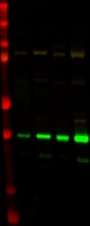
FH Svitlana (Verified Customer) (03-19-2019) | SDS-PAGE: 15 ug protein lysate/well, 4-12% Bis-Tris.Transfer: Immobilon-FL transfer membranes O/N at 30V, 4CBlocking: SEA Block Blocking Buffer 1h at room temperature.Secondary Ab: IRDye 800CW Goat anti-Rabbit 1 h at room temperature.Lines on WB1. BioRad Precision Plus Protein standard2. Homogenate of human cortex tissue3. Non-synaptosomal mitochondria from human cortex4. Homogenate of mice brain tissue5. Non-synaptosomal mitochondria from mice brains
 |

