- Phare
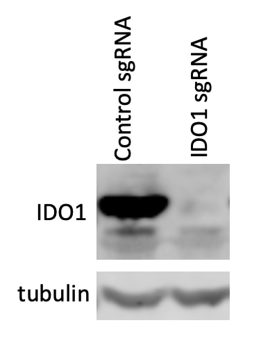
- Validé par KD/KO
Anticorps Polyclonal de lapin anti-IDO1
IDO1 Polyclonal Antibody for WB, IHC, IF/ICC, ELISA
Hôte / Isotype
Lapin / IgG
Réactivité testée
Humain et plus (3)
Applications
WB, IHC, IF/ICC, ELISA
Conjugaison
Non conjugué
N° de cat : 13268-1-AP
Synonymes
Galerie de données de validation
 at dilution of 1:1000 incubated at room temperature for 1.5 hours.")
 at dilution of 1:1000 incubated at room temperature for 1.5 hours.")
 at dilution of 1:200 (under 20x lens). Heat mediated antigen retrieval with Tris-EDTA buffer (pH 9.0).")
 fixed SKOV-3 cells using IDO1 antibody (13268-1-AP) at dilution of 1:200 and CoraLite®488-Conjugated Goat Anti-Rabbit IgG(H+L) (SA00013-2).")
Applications testées
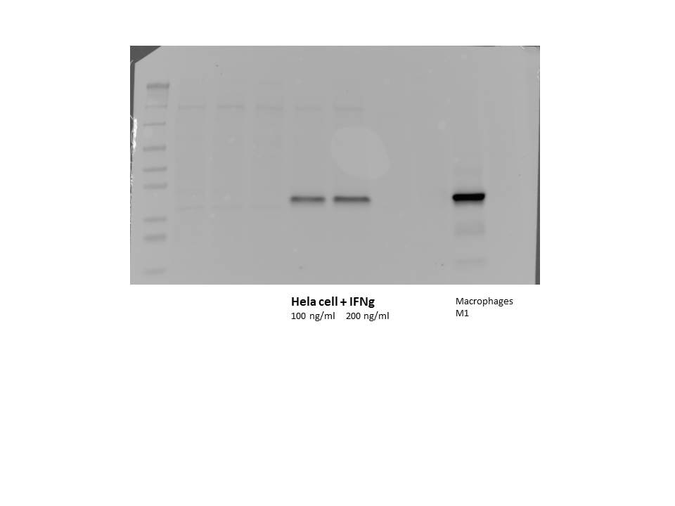
| Résultats positifs en WB | tissu placentaire humain, cellules HeLa traitées à l'IFN gamma |
| Résultats positifs en IHC | human cervical squamous cancer tissue, il est suggéré de démasquer l'antigène avec un tampon de TE buffer pH 9.0; (*) À défaut, 'le démasquage de l'antigène peut être 'effectué avec un tampon citrate pH 6,0. |
| Résultats positifs en IF/ICC | cellules SKOV-3, |
Dilution recommandée
| Application | Dilution |
|---|---|
| Western Blot (WB) | WB : 1:500-1:2000 |
| Immunohistochimie (IHC) | IHC : 1:50-1:500 |
| Immunofluorescence (IF)/ICC | IF/ICC : 1:50-1:500 |
| It is recommended that this reagent should be titrated in each testing system to obtain optimal results. | |
| Sample-dependent, check data in validation data gallery | |
Applications publiées
| KD/KO | See 3 publications below |
| WB | See 70 publications below |
| IHC | See 19 publications below |
| IF | See 15 publications below |
Informations sur le produit
13268-1-AP cible IDO1 dans les applications de WB, IHC, IF/ICC, ELISA et montre une réactivité avec des échantillons Humain
| Réactivité | Humain |
| Réactivité citée | rat, canin, Humain, souris |
| Hôte / Isotype | Lapin / IgG |
| Clonalité | Polyclonal |
| Type | Anticorps |
| Immunogène | IDO1 Protéine recombinante Ag3953 |
| Nom complet | indoleamine 2,3-dioxygenase 1 |
| Masse moléculaire calculée | 403 aa, 45 kDa |
| Poids moléculaire observé | 42 kDa |
| Numéro d’acquisition GenBank | BC027882 |
| Symbole du gène | IDO1 |
| Identification du gène (NCBI) | 3620 |
| Conjugaison | Non conjugué |
| Forme | Liquide |
| Méthode de purification | Purification par affinité contre l'antigène |
| Tampon de stockage | PBS with 0.02% sodium azide and 50% glycerol |
| Conditions de stockage | Stocker à -20°C. Stable pendant un an après l'expédition. L'aliquotage n'est pas nécessaire pour le stockage à -20oC Les 20ul contiennent 0,1% de BSA. |
Informations générales
IDO1 is the target for therapy in a range of clinical settings, including cancer, chronic infections, autoimmune and allergic syndromes, and transplantation. Elevated IDO1 expression is a hallmark of major viral infections including HIV, HBV, HCV or influenza and also of major bacteria infections, such as Tb, CAP, listeriosis and sepsis. Pathogens are able to highjack the immunosuppressive effects of IDO1 and make use of them to facilitate their own life cycle. MW of IDO1 is 40-42kd (PMID: 14502282; 17055065).
Protocole
| Product Specific Protocols | |
|---|---|
| WB protocol for IDO1 antibody 13268-1-AP | Download protocol |
| IHC protocol for IDO1 antibody 13268-1-AP | Download protocol |
| IF protocol for IDO1 antibody 13268-1-AP | Download protocol |
| Standard Protocols | |
|---|---|
| Click here to view our Standard Protocols |
Publications
| Species | Application | Title |
|---|---|---|
Theranostics Single-cell RNA sequencing highlights the immunosuppression of IDO1+ macrophages in the malignant transformation of oral leukoplakia | ||
Hepatology Indoleamine-2, 3-dioxygenase as an effector and an indicator of protective immune responses in patients with acute hepatitis B.
| ||
Acta Biomater Blockage of the IDO1 pathway by charge-switchable nanoparticles amplifies immunogenic cell death for enhanced cancer immunotherapy. | ||
ACS Appl Mater Interfaces Mitigation Effects of Selenium Nanoparticles on Depression-Like Behavior Induced by Fluoride in Mice via the JAK2-STAT3 Pathway. | ||
J Immunother Cancer GCH1 induces immunosuppression through metabolic reprogramming and IDO1 upregulation in triple-negative breast cancer. | ||
J Immunother Cancer Tryptophan potentiates CD8+ T cells against cancer cells by TRIP12 tryptophanylation and surface PD-1 downregulation. |
Avis
The reviews below have been submitted by verified Proteintech customers who received an incentive for providing their feedback.
FH Christopher (Verified Customer) (01-28-2022) | Worked well with a 1 hour incubation at room temperature.
 |
FH Hala (Verified Customer) (09-18-2020) | I am very satisfied of this antibody and of the interaction with your company
 |

