Anticorps Polyclonal de lapin anti-Cytokeratin 10
Cytokeratin 10 Polyclonal Antibody for WB, IHC, IF/ICC, IF-P, FC (Intra), ELISA
Hôte / Isotype
Lapin / IgG
Réactivité testée
Humain, rat, souris
Applications
WB, IHC, IF/ICC, IF-P, FC (Intra), ELISA
Conjugaison
Non conjugué
N° de cat : 18343-1-AP
Synonymes
Galerie de données de validation
 at dilution of 1:200000 incubated at room temperature for 1.5 hours.")
 at dilution of 1:20000 incubated at room temperature for 1.5 hours.")
 at dilution of 1:1000 (under 10x lens). Heat mediated antigen retrieval with Tris-EDTA buffer (pH 9.0).")
 at dilution of 1:200 (under 10x lens. Heat mediated antigen retrieval with Tris-EDTA buffer (pH 9.0).")
 at dilution of 1:200 (under 40x lens. Heat mediated antigen retrieval with Tris-EDTA buffer (pH 9.0).")
 at dilution of 1:1000 (under 40x lens). Heat mediated antigen retrieval with Tris-EDTA buffer (pH 9.0).")
 at dilution of 1:1000 (under 10x lens). Heat mediated antigen retrieval with Tris-EDTA buffer (pH 9.0).")
 at dilution of 1:1000 (under 40x lens). Heat mediated antigen retrieval with Tris-EDTA buffer (pH 9.0).")
 at dilution of 1:200 (under 10x lens).")
 at dilution of 1:200 (under 40x lens).")
 at dilution of 1:200 (under 40x lens).")
 at dilution of 1:200 (under 10x lens).")
 at dilution of 1:200 (under 10x lens).")
 at dilution of 1:200 (under 40x lens).")
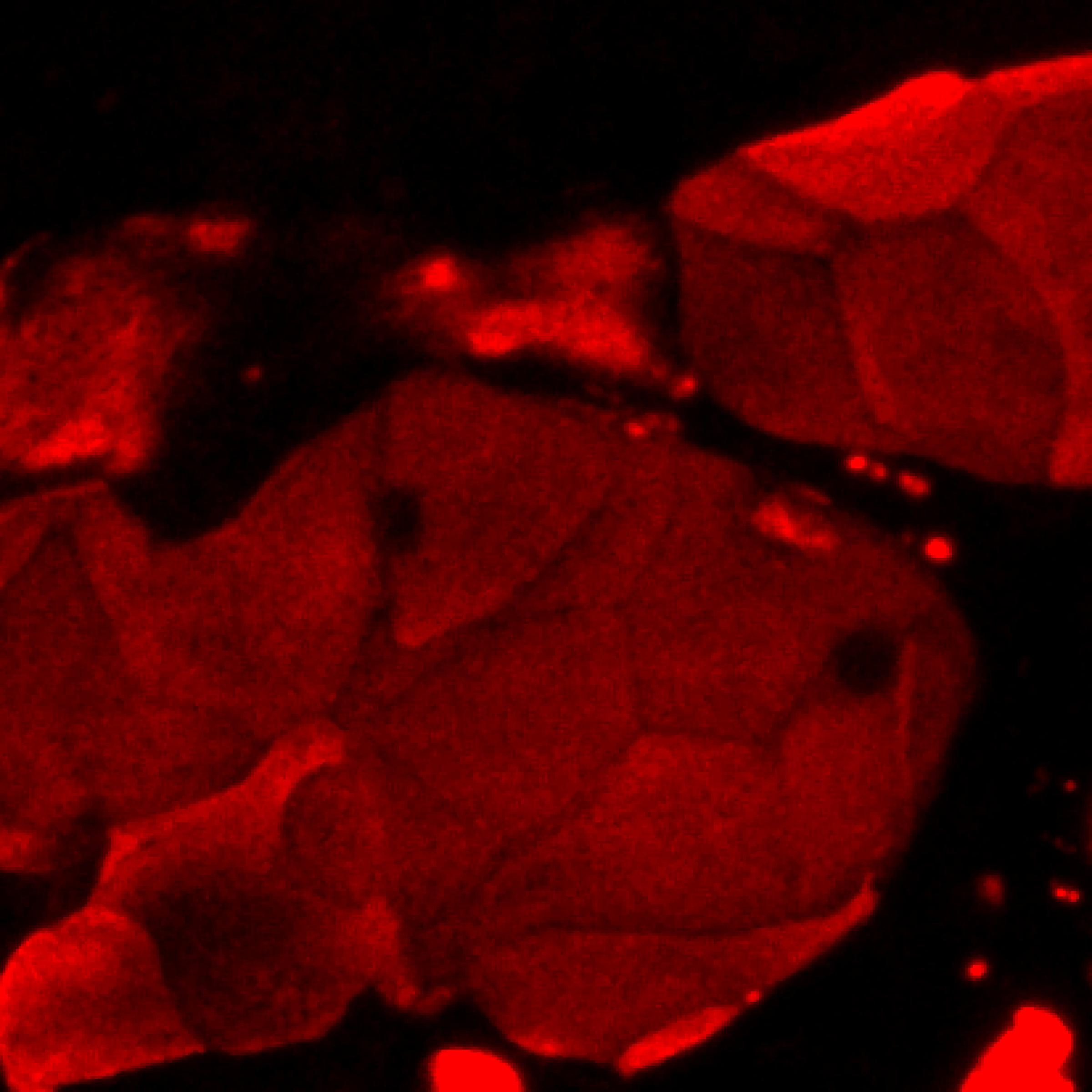
 fixed paraffin-embedded mouse skin tissue using Cytokeratin 10 antibody (18343-1-AP) at dilution of 1:200 and CoraLite®594-Conjugated Goat Anti-Rabbit IgG(H+L) (SA00013-4). Heat mediated antigen retrieval with Tris-EDTA buffer (pH 9.0).")
 fixed A431 cells using Cytokeratin 10 antibody (18343-1-AP) at dilution of 1:200 and CoraLite®488-Conjugated AffiniPure Goat Anti-Rabbit IgG(H+L) (SA00013-2).")
 and CoraLite®488-Conjugated AffiniPure Goat Anti-Rabbit IgG(H+L) at dilution 1:1000 (red), or 0.4 ug rabbit IgG isotype control (blue). Cells were fixed with 4% PFA and permeabilized with Flow Cytometry Perm Buffer (PF00011-C).")
Applications testées
| Résultats positifs en WB | tissu cutané de souris, cellules A431, tissu cutané de rat |
| Résultats positifs en IHC | tissu de cancer du col de l'utérus humain, tissu cutané de rat, tissu de cancer de la peau humain, tissu de cancer du poumon humain, tissu de cancer du sein humain il est suggéré de démasquer l'antigène avec un tampon de TE buffer pH 9.0; (*) À défaut, 'le démasquage de l'antigène peut être 'effectué avec un tampon citrate pH 6,0. |
| Résultats positifs en IF-P | tissu cutané de souris, |
| Résultats positifs en IF/ICC | cellules A431, |
| Résultats positifs en FC (Intra) | cellules A431 |
Dilution recommandée
| Application | Dilution |
|---|---|
| Western Blot (WB) | WB : 1:5000-1:20000 |
| Immunohistochimie (IHC) | IHC : 1:500-1:2000 |
| Immunofluorescence (IF)-P | IF-P : 1:50-1:500 |
| Immunofluorescence (IF)/ICC | IF/ICC : 1:50-1:500 |
| Flow Cytometry (FC) (INTRA) | FC (INTRA) : 0.40 ug per 10^6 cells in a 100 µl suspension |
| It is recommended that this reagent should be titrated in each testing system to obtain optimal results. | |
| Sample-dependent, check data in validation data gallery | |
Applications publiées
| WB | See 3 publications below |
| IHC | See 5 publications below |
| IF | See 11 publications below |
Informations sur le produit
18343-1-AP cible Cytokeratin 10 dans les applications de WB, IHC, IF/ICC, IF-P, FC (Intra), ELISA et montre une réactivité avec des échantillons Humain, rat, souris
| Réactivité | Humain, rat, souris |
| Réactivité citée | rat, Humain, souris |
| Hôte / Isotype | Lapin / IgG |
| Clonalité | Polyclonal |
| Type | Anticorps |
| Immunogène | Cytokeratin 10 Protéine recombinante Ag13136 |
| Nom complet | keratin 10 |
| Masse moléculaire calculée | 584 aa, 59 kDa |
| Poids moléculaire observé | 50-59 kDa |
| Numéro d’acquisition GenBank | BC034697 |
| Symbole du gène | Cytokeratin 10 |
| Identification du gène (NCBI) | 3858 |
| Conjugaison | Non conjugué |
| Forme | Liquide |
| Méthode de purification | Purification par affinité contre l'antigène |
| Tampon de stockage | PBS with 0.02% sodium azide and 50% glycerol |
| Conditions de stockage | Stocker à -20°C. Stable pendant un an après l'expédition. L'aliquotage n'est pas nécessaire pour le stockage à -20oC Les 20ul contiennent 0,1% de BSA. |
Informations générales
Keratins are a large family of proteins that form the intermediate filament cytoskeleton of epithelial cells, which are classified into two major sequence types. Type I keratins are a group of acidic intermediate filament proteins, including K9-K23, and the hair keratins Ha1-Ha8. Type II keratins are the basic or neutral courterparts to the acidic type I keratins, including K1-K8, and the hair keratins, Hb1-Hb6. As a type I keratin, keratin 10 is a suprabasal marker of differentiation in stratified squamous epithelia.
Protocole
| Product Specific Protocols | |
|---|---|
| WB protocol for Cytokeratin 10 antibody 18343-1-AP | Download protocol |
| IHC protocol for Cytokeratin 10 antibody 18343-1-AP | Download protocol |
| IF protocol for Cytokeratin 10 antibody 18343-1-AP | Download protocol |
| FC protocol for Cytokeratin 10 antibody 18343-1-AP | Download protocol |
| Standard Protocols | |
|---|---|
| Click here to view our Standard Protocols |
Publications
| Species | Application | Title |
|---|---|---|
Cell Mol Biol Lett The role of NPY2R/NFATc1/DYRK1A regulatory axis in sebaceous glands for sebum synthesis | ||
Aging Cell Nociceptive transient receptor potential canonical 7 (TRPC7) mediates aging-associated tumorigenesis induced by ultraviolet B. | ||
Life Sci The potential of Diosgenin in treating psoriasis: Studies from HaCaT keratinocytes and imiquimod-induced murine model. | ||
mBio Commensal-Related Changes in the Epidermal Barrier Function Lead to Alterations in the Benzo[a]Pyrene Metabolite Profile and Its Distribution in 3D Skin. | ||
Eur Surg Res A novel combination of keratinocyte and autologous microskin grafting to repair full thickness skin loss | ||
Int J Biol Macromol Skin-adaptive film dressing with smart-release of growth factors accelerated diabetic wound healing |
Avis
The reviews below have been submitted by verified Proteintech customers who received an incentive for providing their feedback.
FH Joshua (Verified Customer) (07-27-2019) | Cells differentiated at air-liquid interface. Cells fixed in 4% paraformaldehyde and stained at 4C overnight. Bright staining with minimal background.
 |

