- Phare
- Validé par KD/KO
Anticorps Polyclonal de lapin anti-LTBP1
LTBP1 Polyclonal Antibody for WB, IHC, IF/ICC, ELISA
Hôte / Isotype
Lapin / IgG
Réactivité testée
Humain et plus (1)
Applications
WB, IHC, IF/ICC, ELISA
Conjugaison
Non conjugué
N° de cat : 26855-1-AP
Synonymes
Galerie de données de validation
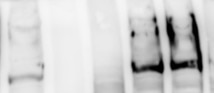
 at dilution of 1:1500 incubated at room temperature for 1.5 hours.")
 at dilution of 1:200 (under 10x lens. Heat mediated antigen retrieval with Tris-EDTA buffer (pH 9.0).")
 at dilution of 1:200 (under 40x lens. Heat mediated antigen retrieval with Tris-EDTA buffer (pH 9.0).")
 fixed HeLa cells using 26855-1-AP (LTBP1 antibody) at dilution of 1:50 and Alexa Fluor 488-Conjugated AffiniPure Goat Anti-Rabbit IgG(H+L).")
Applications testées
| Résultats positifs en WB | tissu plasmatique humain, |
| Résultats positifs en IHC | tissu de cancer du sein humain, il est suggéré de démasquer l'antigène avec un tampon de TE buffer pH 9.0; (*) À défaut, 'le démasquage de l'antigène peut être 'effectué avec un tampon citrate pH 6,0. |
| Résultats positifs en IF/ICC | cellules HeLa, |
Dilution recommandée
| Application | Dilution |
|---|---|
| Western Blot (WB) | WB : 1:500-1:3000 |
| Immunohistochimie (IHC) | IHC : 1:50-1:500 |
| Immunofluorescence (IF)/ICC | IF/ICC : 1:50-1:500 |
| It is recommended that this reagent should be titrated in each testing system to obtain optimal results. | |
| Sample-dependent, check data in validation data gallery | |
Applications publiées
| KD/KO | See 1 publications below |
| WB | See 7 publications below |
| IHC | See 3 publications below |
| IF | See 3 publications below |
Informations sur le produit
26855-1-AP cible LTBP1 dans les applications de WB, IHC, IF/ICC, ELISA et montre une réactivité avec des échantillons Humain
| Réactivité | Humain |
| Réactivité citée | Humain, souris |
| Hôte / Isotype | Lapin / IgG |
| Clonalité | Polyclonal |
| Type | Anticorps |
| Immunogène | LTBP1 Protéine recombinante Ag25392 |
| Nom complet | latent transforming growth factor beta binding protein 1 |
| Masse moléculaire calculée | 1721 aa, 187 kDa |
| Poids moléculaire observé | 150 kDa |
| Numéro d’acquisition GenBank | BC130289 |
| Symbole du gène | LTBP1 |
| Identification du gène (NCBI) | 4052 |
| Conjugaison | Non conjugué |
| Forme | Liquide |
| Méthode de purification | Purification par affinité contre l'antigène |
| Tampon de stockage | PBS with 0.02% sodium azide and 50% glycerol |
| Conditions de stockage | Stocker à -20°C. Stable pendant un an après l'expédition. L'aliquotage n'est pas nécessaire pour le stockage à -20oC Les 20ul contiennent 0,1% de BSA. |
Informations générales
Latent Transforming Growth Factor Beta Binding Protein 1 (LTBP1) is a large extracellular matrix protein that belongs to the latent TGF-beta binding protein family. It plays a crucial role in the regulation of TGF-beta, a multifunctional cytokine involved in various cellular processes such as cell growth, differentiation, and immune response. LTBP1 is essential for TGF-beta folding, secretion, matrix localization, and activation. It forms latent complexes with TGF-beta by covalently binding the TGF-beta propeptide (LAP) via disulfide bonds in the endoplasmic reticulum. This complex is then secreted and targeted to the extracellular matrix, where TGF-beta can be activated by various mechanism.
Protocole
| Product Specific Protocols | |
|---|---|
| WB protocol for LTBP1 antibody 26855-1-AP | Download protocol |
| IHC protocol for LTBP1 antibody 26855-1-AP | Download protocol |
| IF protocol for LTBP1 antibody 26855-1-AP | Download protocol |
| Standard Protocols | |
|---|---|
| Click here to view our Standard Protocols |
Publications
| Species | Application | Title |
|---|---|---|
Cell Rep Integrative single-cell meta-analysis reveals disease-relevant vascular cell states and markers in human atherosclerosis | ||
Phytomedicine Rg3 inhibits hypoxia-induced tumor exosomes from boosting pancreatic cancer vasculogenic mimicry through the HIF-1α/LARS1/mTOR axis | ||
J Transl Med LTBP1 promotes esophageal squamous cell carcinoma progression through epithelial-mesenchymal transition and cancer-associated fibroblasts transformation.
| ||
Cell Death Discov ERRFI1 induces apoptosis of hepatocellular carcinoma cells in response to tryptophan deficiency. | ||
Arterioscler Thromb Vasc Biol Secreted Protein Profiling of Human Aortic Smooth Muscle Cells Identifies Vascular Disease Associations |
Avis
The reviews below have been submitted by verified Proteintech customers who received an incentive for providing their feedback.
FH Udesh (Verified Customer) (10-05-2022) | The Ab was used at 1:1000 dilution in NFDM overnight 4 degrees and detected a band close to 220 kD in 20ug cell lysate. Also tested for for positive IF staining at 1:250 dilution in 2% BSA overnight at 4 degrees.
 |

