Anticorps Polyclonal de lapin anti-MAP2
MAP2 Polyclonal Antibody for WB, IHC, IF/ICC, IF-P, IF-Fro, FC (Intra), IP, ELISA
Hôte / Isotype
Lapin / IgG
Réactivité testée
Humain, rat, souris et plus (2)
Applications
WB, IHC, IF/ICC, IF-P, IF-Fro, FC (Intra), IP, ELISA
Conjugaison
Non conjugué
N° de cat : 17490-1-AP
Synonymes
Galerie de données de validation
 at dilution of 1:30000 incubated at room temperature for 1.5 hours.")
 at dilution of 1:30000 incubated at room temperature for 1.5 hours.")
 at dilution of 1:60000 incubated at room temperature for 1.5 hours.")
 with mouse brain tissue lysate 5000ug.")
 with mouse brain tissue lysate 1280 ug.")
 with SH-SY5Y cells lysate 1240 ug.")
 at dilution of 1:5000 (under 10x lens). Heat mediated antigen retrieval with Tris-EDTA buffer (pH 9.0).")
 at dilution of 1:5000 (under 10x lens). Heat mediated antigen retrieval with Tris-EDTA buffer (pH 9.0).")
 at dilution of 1:5000 (under 40x lens). Heat mediated antigen retrieval with Tris-EDTA buffer (pH 9.0).")
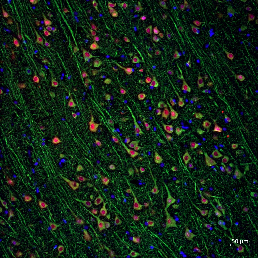
 fixed rat brain tissue using 17490-1-AP (MAP2 antibody) at dilution of 1:100 and CoraLite594-Conjugated AffiniPure Goat Anti-Rabbit IgG(H+L). The section was co-stained with 60190-1-Ig (GFAP antibody, green).")
 fixed rat brain tissue using 17490-1-AP (MAP2 antibody) at dilution of 1:100 and CoraLite594-Conjugated AffiniPure Goat Anti-Rabbit IgG(H+L). The section was co-stained with 60190-1-Ig (GFAP antibody, green).")
 fixed mouse brain tissue using MAP2 antibody (17490-1-AP) at dilution of 1:200 and CoraLite®488-Conjugated AffiniPure Goat Anti-Rabbit IgG(H+L).")
 fixed rat brain tissue using MAP2 antibody (17490-1-AP) at dilution of 1:200 and CoraLite®488-Conjugated AffiniPure Goat Anti-Rabbit IgG(H+L).")
 fixed rat brain tissue using MAP2 antibody (17490-1-AP) at dilution of 1:200 and CoraLite®488-Conjugated AffiniPure Goat Anti-Rabbit IgG(H+L).")
 fixed rat brain tissue using MAP2 antibody (17490-1-AP) at dilution of 1:200 and CoraLite®488-Conjugated AffiniPure Goat Anti-Rabbit IgG(H+L).")
 fixed frozen OCT-embedded mouse brain tissue using MAP2 antibody (17490-1-AP) at dilution of 1:200 and CoraLite®594-Conjugated AffiniPure Goat Anti-Rabbit IgG(H+L) (SA00013-4).")
 fixed frozen OCT-embedded rat brain tissue using MAP2 antibody (17490-1-AP) at dilution of 1:200 and CoraLite®488-Conjugated Goat Anti-Rabbit IgG(H+L) (SA00013-2), GFAP antibody (60190-1-Ig, Clone: 4B2E10, red).")
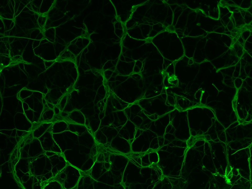
 with 4% PFA fixed control hiPSC derived neuronal cultures (35 days old). (RED MAP2; Blue: DAPI). Provided by BioTalentum Ltd., Hungary.")
 and CoraLite®488-Conjugated AffiniPure Goat Anti-Rabbit IgG(H+L) at dilution 1:1000 (red), or 0.4 ug Control Antibody. Cells were fixed with 4% PFA and permeabilized with Flow Cytometry Perm Buffer (PF00011-C).")
Applications testées
| Résultats positifs en WB | cellules SH-SY5Y, tissu cérébral de rat, tissu cérébral de souris |
| Résultats positifs en IP | cellules SH-SY5Y, tissu cérébral de souris |
| Résultats positifs en IHC | tissu cérébral de souris, il est suggéré de démasquer l'antigène avec un tampon de TE buffer pH 9.0; (*) À défaut, 'le démasquage de l'antigène peut être 'effectué avec un tampon citrate pH 6,0. |
| Résultats positifs en IF-P | tissu cérébral de rat, tissu cérébral de souris |
| Résultats positifs en IF-Fro | tissu cérébral de souris, tissu cérébral de rat |
| Résultats positifs en IF/ICC | cellules iPS, |
| Résultats positifs en FC (Intra) | cellules Neuro-2a |
Dilution recommandée
| Application | Dilution |
|---|---|
| Western Blot (WB) | WB : 1:5000-1:50000 |
| Immunoprécipitation (IP) | IP : 0.5-4.0 ug for 1.0-3.0 mg of total protein lysate |
| Immunohistochimie (IHC) | IHC : 1:2500-1:10000 |
| Immunofluorescence (IF)-P | IF-P : 1:50-1:500 |
| Immunofluorescence (IF)-FRO | IF-FRO : 1:50-1:500 |
| Immunofluorescence (IF)/ICC | IF/ICC : 1:125-1:500 |
| Flow Cytometry (FC) (INTRA) | FC (INTRA) : 0.40 ug per 10^6 cells in a 100 µl suspension |
| It is recommended that this reagent should be titrated in each testing system to obtain optimal results. | |
| Sample-dependent, check data in validation data gallery | |
Applications publiées
| WB | See 89 publications below |
| IHC | See 35 publications below |
| IF | See 222 publications below |
Informations sur le produit
17490-1-AP cible MAP2 dans les applications de WB, IHC, IF/ICC, IF-P, IF-Fro, FC (Intra), IP, ELISA et montre une réactivité avec des échantillons Humain, rat, souris
| Réactivité | Humain, rat, souris |
| Réactivité citée | rat, Chèvre, Humain, singe, souris |
| Hôte / Isotype | Lapin / IgG |
| Clonalité | Polyclonal |
| Type | Anticorps |
| Immunogène | MAP2 Protéine recombinante Ag11580 |
| Nom complet | microtubule-associated protein 2 |
| Masse moléculaire calculée | 200 kDa |
| Poids moléculaire observé | 280 kDa, 70-85 kDa |
| Numéro d’acquisition GenBank | BC038857 |
| Symbole du gène | MAP2 |
| Identification du gène (NCBI) | 4133 |
| Conjugaison | Non conjugué |
| Forme | Liquide |
| Méthode de purification | Purification par affinité contre l'antigène |
| Tampon de stockage | PBS with 0.02% sodium azide and 50% glycerol |
| Conditions de stockage | Stocker à -20°C. Stable pendant un an après l'expédition. L'aliquotage n'est pas nécessaire pour le stockage à -20oC Les 20ul contiennent 0,1% de BSA. |
Informations générales
Microtubule-associated protein 2 (MAP2) is a tubulin binding protein regulating the spacing and stability of microtubules and contributing to elongation of dendrites.
What is the molecular weight of MAP2? Is MAP2 post-translationally modified?
MAP2 has multiple isoforms that arise from alternative splicing (PMID: 3121794, 7854050, and 10383434). They are classified into two groups - MAP2A and MAP2B, which are known as high molecular weight (HMW) isoforms, run as ~280 kDa species, while low molecular weight (LMW) isoforms MAP2C and MAP2D are around ~70 kDa. MAP2 proteins are heavily phosphorylated, which contributes to a large discrepancy between their predicted and observed molecular weight in SDS-PAGE (220 vs 280 kDa for HMW forms).
What is the tissue expression pattern of MAP2? What is the subcellular localization of MAP2?
MAP2 isoforms differ in their tissue and developmental expression pattern (PMID: 2469170 and 3898077). In the brain, MAP2B is widely expressed during and post development, MAP2A is expressed postnatally, while MAP2C is present only in the early development except of present in photosensitive cells of the adult retina and in the olfactory system. MAP proteins are highly expressed in the CNS found in cell bodies and dendrites of neurons, in dorsal root ganglion, reactive glia, and in the testis (PMID: 9588626). In neurons, MAP2 proteins are found in the cell body and dendrites, where they associate with microtubules, while they can also be present in the nuclei of testicular cells.
Can MAP2 be used as a neuronal marker?
MAP2 proteins are abundantly expressed in neurons. MAP2 is frequently used as a dendritic marker because it is present in the cell body and dendrites of neurons but absent in axons (PMID: 28413822).
Protocole
| Product Specific Protocols | |
|---|---|
| WB protocol for MAP2 antibody 17490-1-AP | Download protocol |
| IHC protocol for MAP2 antibody 17490-1-AP | Download protocol |
| IF protocol for MAP2 antibody 17490-1-AP | Download protocol |
| IP protocol for MAP2 antibody 17490-1-AP | Download protocol |
| Standard Protocols | |
|---|---|
| Click here to view our Standard Protocols |
Publications
| Species | Application | Title |
|---|---|---|
Cell Stem Cell In vivo reprogramming of NG2 glia enables adult neurogenesis and functional recovery following spinal cord injury. | ||
Cell Stem Cell Recent Zika Virus Isolates Induce Premature Differentiation of Neural Progenitors in Human Brain Organoids. | ||
Nat Commun Reliability of high-quantity human brain organoids for modeling microcephaly, glioma invasion and drug screening | ||
Nat Commun Repair-associated macrophages increase after early-phase microglia attenuation to promote ischemic stroke recovery | ||
Neuron Astrocytic ApoE reprograms neuronal cholesterol metabolism and histone-acetylation-mediated memory. | ||
Sci Adv Glioblastoma exploits ATP from leading-edge astrocytes to fuel its infiltrative growth revealed by spatially resolved chimeric analysis |
Avis
The reviews below have been submitted by verified Proteintech customers who received an incentive for providing their feedback.
FH Makenna (Verified Customer) (07-15-2024) | Antibody worked well for IF at a dilution of 1:500
|
FH Abigail (Verified Customer) (07-15-2024) | Used for IF and got clear images in green and red channels.
|
FH Reyes (Verified Customer) (04-05-2024) | MAP2 (in green) shows a strong marking of my neurons in human brain FFPE cortex
 |
FH Filipa (Verified Customer) (09-18-2023) | I used this anti-MAP2 antibody at 1:200 dilution for IF. The cells were fixed in 4% PFA in PBS for 20 minutes, and incubated with the primary antibody for 3 hours at RT. MAP2 staining works very well at this dilution.
 |
FH BING (Verified Customer) (08-04-2021) | suitable for detection of both bands of MAP2 by WB with one above 250 kd
 |
FH Dipen (Verified Customer) (07-20-2020) | Not great for Western blotting.
 |
FH Naomi (Verified Customer) (03-09-2020) | I used the rabbit polyclonal anti-MAP2 antibody at 1:500 dilution to in differentiated PC-12 cells. The cells were fixed in 4% PFA in PBS for 12 minutes. 1 hr in blocking buffer, followed by 24 h incubation in primary antibody. MAP2 was expressed in neurites. Works well at this dilution, great images.
|
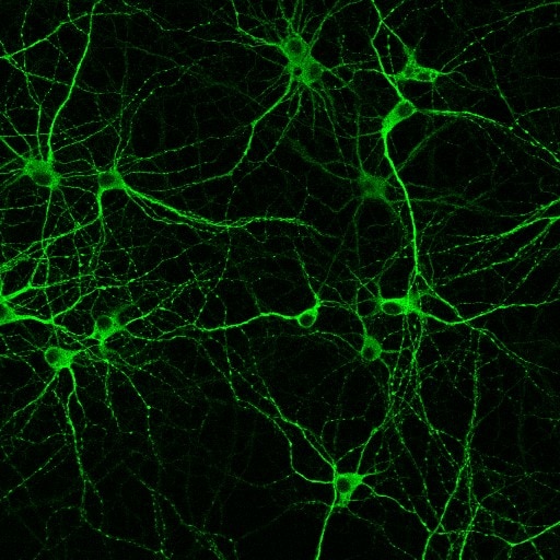
FH Gloria (Verified Customer) (01-30-2020) | Beautiful staining in primary hippocampal and cortical neurons.
 |
FH Ricky (Verified Customer) (02-22-2019) | 1:5000 dilution is good enough for a clear and sharp image of western blot. I am now using Li-Cor's Odyssey CLX imaging systems . The second antibody is from Li-Cor with 1:5000 dilution. By the way, I also tried to use 1:1000 dilution for my low expression samples, and there was no bad noise backgrounds.
|

