- Phare
- Validé par KD/KO
Anticorps Polyclonal de lapin anti-MAP3K1
MAP3K1 Polyclonal Antibody for WB, IHC, IF/ICC, IP, ELISA
Hôte / Isotype
Lapin / IgG
Réactivité testée
Humain, rat, souris
Applications
WB, IHC, IF/ICC, IP, ELISA
Conjugaison
Non conjugué
N° de cat : 19970-1-AP
Synonymes
Galerie de données de validation
Applications testées
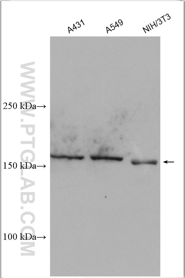
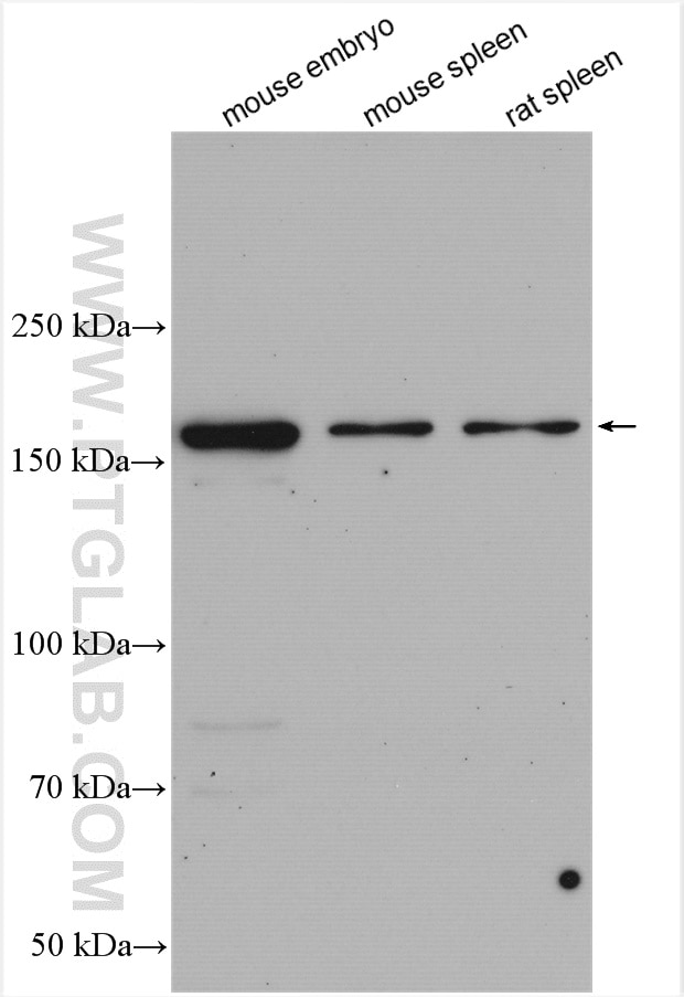
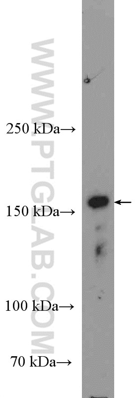
| Résultats positifs en WB | tissu embryonnaire de souris, cellules A431, cellules A549, cellules NIH/3T3, tissu splénique de rat, tissu splénique de souris |
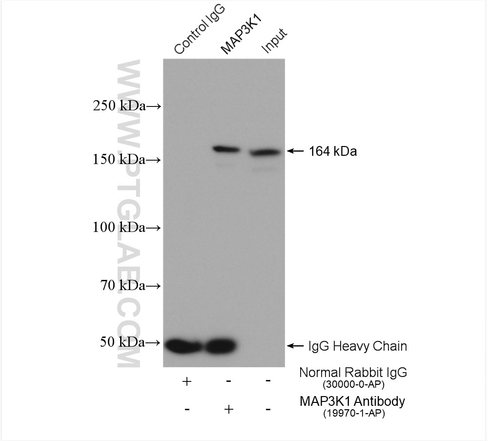
| Résultats positifs en IP | cellules NIH/3T3, |
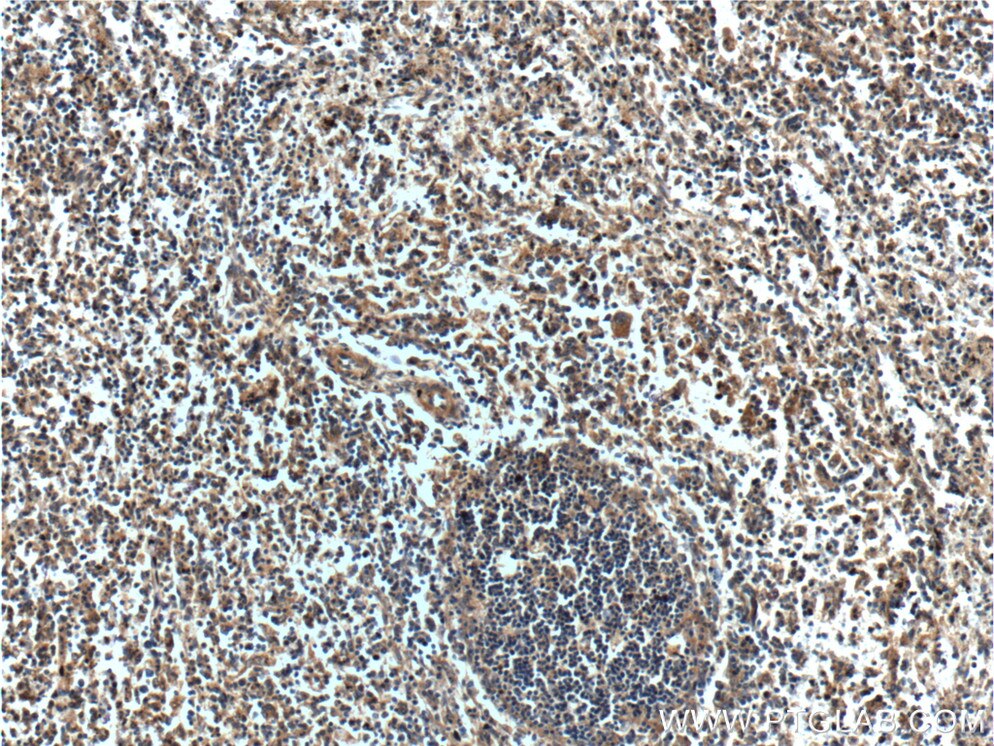
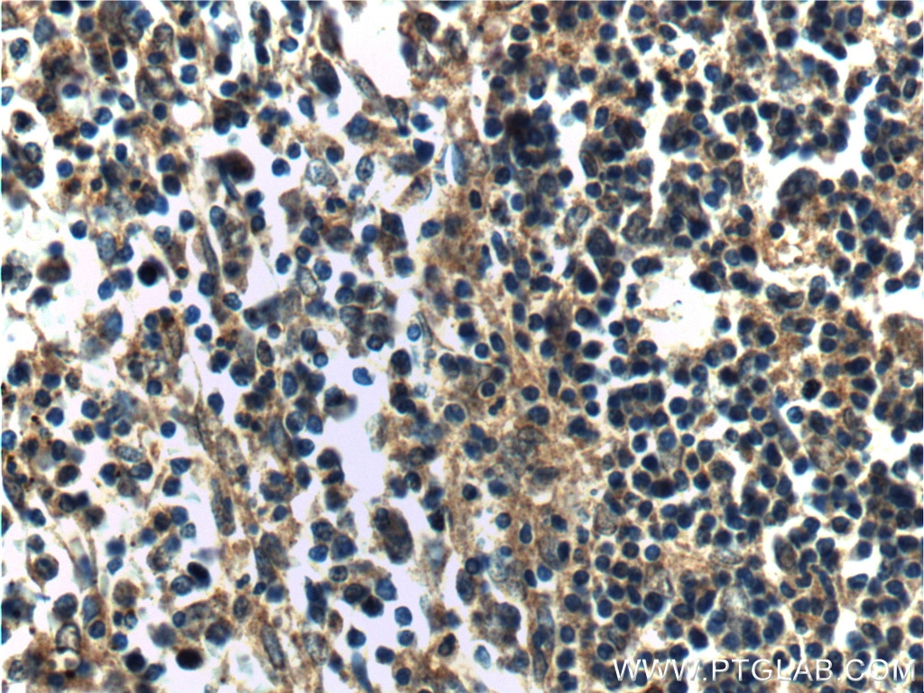
| Résultats positifs en IHC | tissu splénique humain il est suggéré de démasquer l'antigène avec un tampon de TE buffer pH 9.0; (*) À défaut, 'le démasquage de l'antigène peut être 'effectué avec un tampon citrate pH 6,0. |
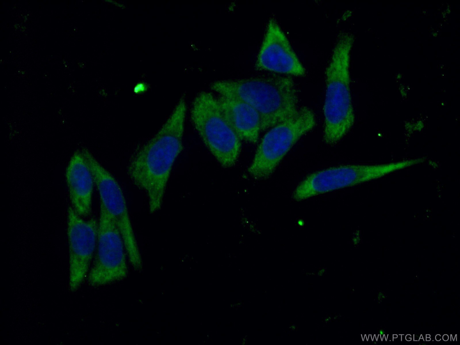
| Résultats positifs en IF/ICC | cellules HeLa |
Dilution recommandée
| Application | Dilution |
|---|---|
| Western Blot (WB) | WB : 1:1000-1:4000 |
| Immunoprécipitation (IP) | IP : 0.5-4.0 ug for 1.0-3.0 mg of total protein lysate |
| Immunohistochimie (IHC) | IHC : 1:50-1:500 |
| Immunofluorescence (IF)/ICC | IF/ICC : 1:20-1:200 |
| It is recommended that this reagent should be titrated in each testing system to obtain optimal results. | |
| Sample-dependent, check data in validation data gallery | |
Applications publiées
| KD/KO | See 2 publications below |
| WB | See 12 publications below |
| IF | See 2 publications below |
Informations sur le produit
19970-1-AP cible MAP3K1 dans les applications de WB, IHC, IF/ICC, IP, ELISA et montre une réactivité avec des échantillons Humain, rat, souris
| Réactivité | Humain, rat, souris |
| Réactivité citée | Humain, souris |
| Hôte / Isotype | Lapin / IgG |
| Clonalité | Polyclonal |
| Type | Anticorps |
| Immunogène | Peptide |
| Nom complet | mitogen-activated protein kinase kinase kinase 1 |
| Masse moléculaire calculée | 164 kDa |
| Poids moléculaire observé | 164 kDa |
| Numéro d’acquisition GenBank | NM_005921 |
| Symbole du gène | MAP3K1 |
| Identification du gène (NCBI) | 4214 |
| Conjugaison | Non conjugué |
| Forme | Liquide |
| Méthode de purification | Purification par affinité contre l'antigène |
| Tampon de stockage | PBS with 0.02% sodium azide and 50% glycerol |
| Conditions de stockage | Stocker à -20°C. Stable pendant un an après l'expédition. L'aliquotage n'est pas nécessaire pour le stockage à -20oC Les 20ul contiennent 0,1% de BSA. |
Informations générales
MAP3K1, also named as MAPKKK1, MEKK and MEKK1, belongs to the protein kinase superfamily, STE Ser/Thr protein kinase family and MAP kinase kinase kinase subfamily. MAP3K1 is a component of a protein kinase signal transduction cascade. MAP3K1 activates the ERK and JNK kinase pathways by phosphorylation of MAP2K1 and MAP2K4. It activates CHUK and IKBKB, the central protein kinases of the NF-kappa-B pathway. It catalyzes the reaction: ATP + a protein = ADP + a phosphoprotein. The antibody recognizes the N-term of MAP3K1.
Protocole
| Product Specific Protocols | |
|---|---|
| WB protocol for MAP3K1 antibody 19970-1-AP | Download protocol |
| IHC protocol for MAP3K1 antibody 19970-1-AP | Download protocol |
| IF protocol for MAP3K1 antibody 19970-1-AP | Download protocol |
| IP protocol for MAP3K1 antibody 19970-1-AP | Download protocol |
| Standard Protocols | |
|---|---|
| Click here to view our Standard Protocols |
Publications
| Species | Application | Title |
|---|---|---|
J Immunol Res Circulating miR-320b Contributes to CD4+ T-Cell Proliferation in Systemic Lupus Erythematosus via MAP3K1 | ||
Int J Biol Sci LncRNA SLCO4A1-AS1 predicts poor prognosis and promotes proliferation and metastasis via the EGFR/MAPK pathway in colorectal cancer. | ||
Int Immunopharmacol Extracellular vesicles secreted from mesenchymal stem cells exert anti-apoptotic and anti-inflammatory effects via transmitting microRNA-18b in rats with diabetic retinopathy. | ||
Biomed Pharmacother Xihuang pill promotes apoptosis of Treg cells in the tumor microenvironment in 4T1 mouse breast cancer by upregulating MEKK1/SEK1/JNK1/AP-1 pathway. | ||
Cell Death Dis LncRNA KCNQ1OT1 activated by c-Myc promotes cell proliferation via interacting with FUS to stabilize MAP3K1 in acute promyelocytic leukemia. | ||
Biomed Res Int Next-Generation Sequencing Panel Analysis of Clinically Relevant Mutations in Circulating Cell-Free DNA from Patients with Gestational Trophoblastic Neoplasia: A Pilot Study. |
