- Phare
- Validé par KD/KO
Anticorps Polyclonal de lapin anti-MCT1
MCT1 Polyclonal Antibody for WB, IHC, IF/ICC, IP, ELISA
Hôte / Isotype
Lapin / IgG
Réactivité testée
Humain et plus (3)
Applications
WB, IHC, IF/ICC, IP, ELISA
Conjugaison
Non conjugué
N° de cat : 20139-1-AP
Synonymes
Galerie de données de validation
 with sh-Control and sh-MCT1 transfected HeLa cells.")
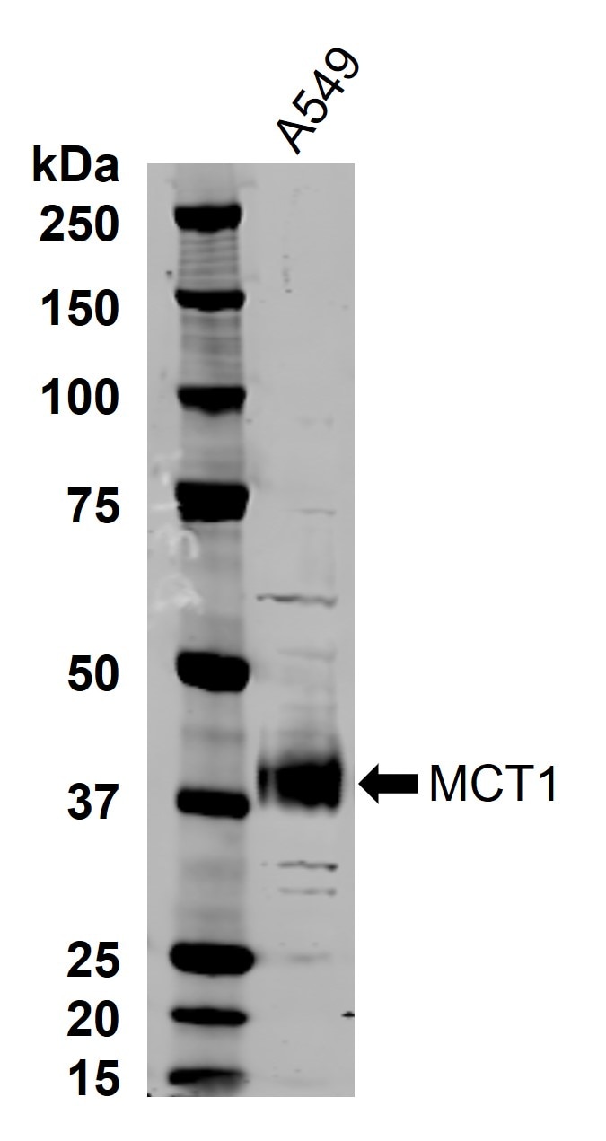
 at dilution of 1:5000 incubated at room temperature for 1.5 hours.")
 with Raji cells lysate 3600 ug.")
 at dilution of 1:600 (under 10x lens. Heat mediated antigen retrieval with Tris-EDTA buffer (pH 9.0).")
 at dilution of 1:600 (under 40x lens. Heat mediated antigen retrieval with Tris-EDTA buffer (pH 9.0).")
 at dilution of 1:1600 (under 20x lens). Heat mediated antigen retrieval with Tris-EDTA buffer (pH 9.0).")
 at dilution of 1:400 (under 10x lens. Heat mediated antigen retrieval with Tris-EDTA buffer (pH 9.0).")
 at dilution of 1:400 (under 40x lens. Heat mediated antigen retrieval with Tris-EDTA buffer (pH 9.0).")
 at dilution of 1:800 (under 10x lens). Heat mediated antigen retrieval with Tris-EDTA buffer (pH 9.0).")
 at dilution of 1:800 (under 40x lens). Heat mediated antigen retrieval with Tris-EDTA buffer (pH 9.0).")
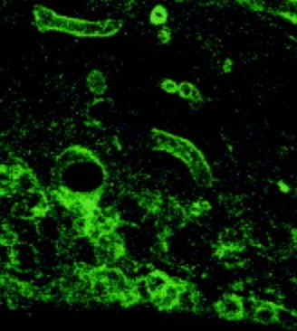
 fixed HeLa cells using MCT1 antibody (20139-1-AP) at dilution of 1:200 and CoraLite®594-Conjugated Goat Anti-Rabbit IgG(H+L) (SA00013-4).")
Applications testées
| Résultats positifs en WB | cellules BxPC-3, cellules HeLa |
| Résultats positifs en IP | cellules Raji, |
| Résultats positifs en IHC | tissu de cancer du côlon humain, tissu de cancer du col de l'utérus humain, tissu de cancer du sein humain il est suggéré de démasquer l'antigène avec un tampon de TE buffer pH 9.0; (*) À défaut, 'le démasquage de l'antigène peut être 'effectué avec un tampon citrate pH 6,0. |
| Résultats positifs en IF/ICC | cellules HeLa, |
Dilution recommandée
| Application | Dilution |
|---|---|
| Western Blot (WB) | WB : 1:2000-1:10000 |
| Immunoprécipitation (IP) | IP : 0.5-4.0 ug for 1.0-3.0 mg of total protein lysate |
| Immunohistochimie (IHC) | IHC : 1:600-1:2000 |
| Immunofluorescence (IF)/ICC | IF/ICC : 1:50-1:500 |
| It is recommended that this reagent should be titrated in each testing system to obtain optimal results. | |
| Sample-dependent, check data in validation data gallery | |
Applications publiées
| KD/KO | See 8 publications below |
| WB | See 69 publications below |
| IHC | See 28 publications below |
| IF | See 26 publications below |
| IP | See 1 publications below |
Informations sur le produit
20139-1-AP cible MCT1 dans les applications de WB, IHC, IF/ICC, IP, ELISA et montre une réactivité avec des échantillons Humain
| Réactivité | Humain |
| Réactivité citée | rat, bovin, Humain, souris |
| Hôte / Isotype | Lapin / IgG |
| Clonalité | Polyclonal |
| Type | Anticorps |
| Immunogène | MCT1 Protéine recombinante Ag14098 |
| Nom complet | solute carrier family 16, member 1 (monocarboxylic acid transporter 1) |
| Masse moléculaire calculée | 500 aa, 54 kDa |
| Poids moléculaire observé | 38-45 kDa |
| Numéro d’acquisition GenBank | BC026317 |
| Symbole du gène | MCT1 |
| Identification du gène (NCBI) | 6566 |
| Conjugaison | Non conjugué |
| Forme | Liquide |
| Méthode de purification | Purification par affinité contre l'antigène |
| Tampon de stockage | PBS with 0.02% sodium azide and 50% glycerol |
| Conditions de stockage | Stocker à -20°C. Stable pendant un an après l'expédition. L'aliquotage n'est pas nécessaire pour le stockage à -20oC Les 20ul contiennent 0,1% de BSA. |
Informations générales
MCT1, the SLC16A1 gene product, is a trans-membrane symporter involved in lactate and pyruvate transportation. It plays an important role in lactic acid transport and H+ clearance in cancer cells. MCT overexpression had been observed in various cancers and may play an important role in tumorigenesis. Two isoforms of MCT1 exist due to the alternative splicing, with predicted MW of 54 kDa and 46 kDa, respectively. While western blot analysis detected MCT1 at an apparent molecular mass of 40-50 kDa.
Protocole
| Product Specific Protocols | |
|---|---|
| WB protocol for MCT1 antibody 20139-1-AP | Download protocol |
| IHC protocol for MCT1 antibody 20139-1-AP | Download protocol |
| IF protocol for MCT1 antibody 20139-1-AP | Download protocol |
| IP protocol for MCT1 antibody 20139-1-AP | Download protocol |
| Standard Protocols | |
|---|---|
| Click here to view our Standard Protocols |
Publications
| Species | Application | Title |
|---|---|---|
Cell Metab High dietary fructose promotes hepatocellular carcinoma progression by enhancing O-GlcNAcylation via microbiota-derived acetate | ||
Cell Metab Acetate enables metabolic fitness and cognitive performance during sleep disruption | ||
Nat Cancer Targeting the bicarbonate transporter SLC4A4 overcomes immunosuppression and immunotherapy resistance in pancreatic cancer | ||
J Exp Clin Cancer Res Lnc AC016727.1/BACH1/HIF-1 α signal loop promotes the progression of non-small cell lung cancer | ||
Clin Cancer Res A Novel Role for DNA-PK in Metabolism by Regulating Glycolysis in Castration Resistant Prostate Cancer. | ||
Theranostics ITGB2-mediated metabolic switch in CAFs promotes OSCC proliferation by oxidation of NADH in mitochondrial oxidative phosphorylation system. |
Avis
The reviews below have been submitted by verified Proteintech customers who received an incentive for providing their feedback.
FH Angie (Verified Customer) (08-27-2024) | A549 lysate was subjected to western blot with MCT1 antibody used at 1:10000 dilution and incubated at room temperature for 1.5 hours. Secondary antibody Donkey-anti-rabbit (Alexa Fluor 800) used at 1:20000 dilution incubated at room temperature for 1 hour. One thick band at around 40 kDa. Image is shown in grayscale.
 |
FH Xie (Verified Customer) (07-07-2023) | The expression of MCT4 was seen in my samples.
 |
FH Aderonke (Verified Customer) (05-23-2023) | The expression of MCT4 was seen in three of the five cell lines used without an inhibitor. Also, an inhibtor of MCT4, was used at two different concentrations and the expression was decreased in the cells that expressed the protein.
|
FH Huseyin (Verified Customer) (07-28-2022) | I used the antibody with 1:3000 dilution in TBS-T solution O/N and I received correct size bands in Ishikawa cell line.
|

