- Phare
- Validé par KD/KO
Anticorps Polyclonal de lapin anti-MICAL1
MICAL1 Polyclonal Antibody for WB, IHC, IF/ICC, IP, ELISA
Hôte / Isotype
Lapin / IgG
Réactivité testée
Humain et plus (1)
Applications
WB, IHC, IF/ICC, IP, CoIP, ELISA
Conjugaison
Non conjugué
N° de cat : 14818-1-AP
Synonymes
Galerie de données de validation
 with sh-Control and sh-MICAL1 transfected HeLa cells.")
 at dilution of 1:8000 incubated at room temperature for 1.5 hours.")
 at dilution of 1:500 incubated at room temperature for 1.5 hours.")
 at dilution of 1:1000 incubated at room temperature for 1.5 hours.")
 at dilution of 1:500 incubated at room temperature for 1.5 hours.")
 at dilution of 1:500 incubated at room temperature for 1.5 hours.")
 at dilution of 1:1000 incubated at room temperature for 1.5 hours.")
 with HeLa cells lysate 2000ug.")
 with T-47D cells lysate 1800 ug.")
 at dilution of 1:100 (under 10x lens).")
 at dilution of 1:100 (under 40x lens).")
 at dilution of 1:100 (under 10x lens).")
 at dilution of 1:100 (under 40x lens).")
 at dilution of 1:100 (under 10x lens).")
 at dilution of 1:100 (under 40x lens).")
 at dilution of 1:100 (under 10x lens).")
 at dilution of 1:100 (under 40x lens).")
 at dilution of 1:100 (under 10x lens).")
 at dilution of 1:100 (under 40x lens).")
 at dilution of 1:100 (under 10x lens).")
 at dilution of 1:100 (under 40x lens).")
 at dilution of 1:100 (under 10x lens).")
 at dilution of 1:100 (under 40x lens).")
 fixed A549 cells using MICAL1 antibody (14818-1-AP) at dilution of 1:400 and CoraLite®488-Conjugated AffiniPure Goat Anti-Rabbit IgG(H+L), Alpha Tubulin antibody (66031-1-Ig, Clone: 1E4C11, red).")
 fixed HeLa cells using 14818-1-AP (MICAL1 antibody) at dilution of 1:50 and Alexa Fluor 488-Conjugated AffiniPure Goat Anti-Rabbit IgG(H+L).")
 with Hela Cells (Methanol fixation) MICAL1 in Red (endogenous) and BioGFP-MICAL1-FL overexpression. Courtesy of Qingyang Liu, Utrecht University.")
Applications testées
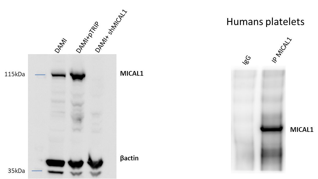
| Résultats positifs en WB | cellules Jurkat, cellules HEK-293, cellules HeLa, cellules T-47D, tissu cérébral humain |
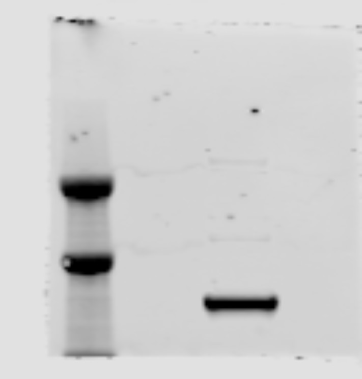
| Résultats positifs en IP | cellules HeLa, cellules T-47D |
| Résultats positifs en IHC | tissu pulmonaire humain, tissu cardiaque humain, tissu cutané humain, tissu ovarien humain, tissu placentaire humain, tissu splénique humain, tissu testiculaire humain il est suggéré de démasquer l'antigène avec un tampon de TE buffer pH 9.0; (*) À défaut, 'le démasquage de l'antigène peut être 'effectué avec un tampon citrate pH 6,0. |
| Résultats positifs en IF/ICC | cellules A549, cellules HeLa |
Dilution recommandée
| Application | Dilution |
|---|---|
| Western Blot (WB) | WB : 1:2000-1:16000 |
| Immunoprécipitation (IP) | IP : 0.5-4.0 ug for 1.0-3.0 mg of total protein lysate |
| Immunohistochimie (IHC) | IHC : 1:20-1:200 |
| Immunofluorescence (IF)/ICC | IF/ICC : 1:200-1:800 |
| It is recommended that this reagent should be titrated in each testing system to obtain optimal results. | |
| Sample-dependent, check data in validation data gallery | |
Applications publiées
| KD/KO | See 11 publications below |
| WB | See 16 publications below |
| IHC | See 6 publications below |
| IF | See 9 publications below |
| IP | See 1 publications below |
| CoIP | See 2 publications below |
Informations sur le produit
14818-1-AP cible MICAL1 dans les applications de WB, IHC, IF/ICC, IP, CoIP, ELISA et montre une réactivité avec des échantillons Humain
| Réactivité | Humain |
| Réactivité citée | Humain, souris |
| Hôte / Isotype | Lapin / IgG |
| Clonalité | Polyclonal |
| Type | Anticorps |
| Immunogène | MICAL1 Protéine recombinante Ag6578 |
| Nom complet | microtubule associated monoxygenase, calponin and LIM domain containing 1 |
| Masse moléculaire calculée | 118 kDa |
| Poids moléculaire observé | 120 kDa |
| Numéro d’acquisition GenBank | BC052983 |
| Symbole du gène | MICAL1 |
| Identification du gène (NCBI) | 64780 |
| Conjugaison | Non conjugué |
| Forme | Liquide |
| Méthode de purification | Purification par affinité contre l'antigène |
| Tampon de stockage | PBS with 0.02% sodium azide and 50% glycerol |
| Conditions de stockage | Stocker à -20°C. Stable pendant un an après l'expédition. L'aliquotage n'est pas nécessaire pour le stockage à -20oC Les 20ul contiennent 0,1% de BSA. |
Informations générales
MICALs (Molecules Interacting with CasL) are atypical multidomain flavoenzymes with diverse cellular functions.There are three known isoforms, MICAL1, MICAL2 and MICAL3, as well as the MICAL-like proteins MICAL-L1 and MICAL-L2. MICAL1 has four conserved domains: an N-terminal flavin adenine dinucleotide (FAD) binding domain, a calponin homology (CH) domain, a Lin11, Isl-1 and Mec-3 (LIM) domain and a C-terminal coiled-coil (CC) domain. MICAL1 is reported to regulate actin stress fibers and be required for normal actin organization. It may also be involved in apoptosis through binding with NDR (nuclear Dbf2-related) kinases. This antibody specially recognizes MICAL1.
Protocole
| Product Specific Protocols | |
|---|---|
| WB protocol for MICAL1 antibody 14818-1-AP | Download protocol |
| IHC protocol for MICAL1 antibody 14818-1-AP | Download protocol |
| IF protocol for MICAL1 antibody 14818-1-AP | Download protocol |
| IP protocol for MICAL1 antibody 14818-1-AP | Download protocol |
| Standard Protocols | |
|---|---|
| Click here to view our Standard Protocols |
Publications
| Species | Application | Title |
|---|---|---|
Sci Adv F-actin disassembly factor MICAL1 binding to Myosin Va mediates cargo unloading during cytokinesis.
| ||
Dev Cell Amplification of F-Actin Disassembly and Cellular Repulsion by Growth Factor Signaling. | ||
J Cell Biol MICAL2 enhances branched actin network disassembly by oxidizing Arp3B-containing Arp2/3 complexes.
| ||
Oncogene Phosphorylation of MICAL2 by ARG promotes head and neck cancer tumorigenesis by regulating skeletal rearrangement | ||
Avis
The reviews below have been submitted by verified Proteintech customers who received an incentive for providing their feedback.
FH Kincaid (Verified Customer) (06-02-2024) | Tested antibody in H4 cells. Diluted aMICAL1 1:500 in 3% BSA, diluted in TBS-tween (1%).
 |
FH Christelle (Verified Customer) (11-21-2023) | For wb: used dilution 1/1000 For IP: used 2µg of antibody
 |
FH Joleen (Verified Customer) (06-03-2019) | Shows band at where MICAL1 is expected but is faint. This image is a merge image of the membrane exposed for 120s and a stain free image to visualize the molecular weight. Even with long exposure, the band is faint.
 |

