- Phare
- Validé par KD/KO
Anticorps Polyclonal de lapin anti-MIF
MIF Polyclonal Antibody for WB, IF/ICC, FC (Intra), IP, ELISA
Hôte / Isotype
Lapin / IgG
Réactivité testée
Humain, rat, souris
Applications
WB, IF/ICC, FC (Intra), IP, ELISA
Conjugaison
Non conjugué
N° de cat : 20415-1-AP
Synonymes
Galerie de données de validation
 at dilution of 1:1000 incubated at room temperature for 1.5 hours.")
 at dilution of 1:1000 incubated at room temperature for 1.5 hours.")
 at dilution of 1:15000 incubated at room temperature for 1.5 hours.")
 at dilution of 1:10000 incubated at room temperature for 1.5 hours.")
 at dilution of 1:1000 incubated at room temperature for 1.5 hours.")
 at dilution of 1:1000 incubated at room temperature for 1.5 hours.")
 at dilution of 1:500 incubated at room temperature for 1.5 hours.")
 with mouse spleen tissue lysate 4000ug.")
 fixed U-251 cells using MIF antibody (20415-1-AP) at dilution of 1:200 and CoraLite®488-Conjugated AffiniPure Goat Anti-Rabbit IgG(H+L).")
 and CoraLite®488-Conjugated AffiniPure Goat Anti-Rabbit IgG(H+L) at dilution 1:1000 (red), or 0.4 ug Control Antibody. Cells were fixed with 4% PFA and permeabilized with Flow Cytometry Perm Buffer (PF00011-C).")
Applications testées
| Résultats positifs en WB | cellules A549, cellules HEK-293T, cellules HL-60, cellules HUVEC, cellules Jurkat, cellules MG U-87, cellules Neuro-2a, cellules U-937, cellules Y79, tissu splénique de rat, tissu splénique de souris |
| Résultats positifs en IP | tissu splénique de souris |
| Résultats positifs en IF/ICC | cellules U-251, |
| Résultats positifs en FC (Intra) | cellules THP-1 |
Dilution recommandée
| Application | Dilution |
|---|---|
| Western Blot (WB) | WB : 1:500-1:2000 |
| Immunoprécipitation (IP) | IP : 0.5-4.0 ug for 1.0-3.0 mg of total protein lysate |
| Immunofluorescence (IF)/ICC | IF/ICC : 1:50-1:500 |
| Flow Cytometry (FC) (INTRA) | FC (INTRA) : 0.40 ug per 10^6 cells in a 100 µl suspension |
| It is recommended that this reagent should be titrated in each testing system to obtain optimal results. | |
| Sample-dependent, check data in validation data gallery | |
Applications publiées
| KD/KO | See 6 publications below |
| WB | See 16 publications below |
| IF | See 9 publications below |
Informations sur le produit
20415-1-AP cible MIF dans les applications de WB, IF/ICC, FC (Intra), IP, ELISA et montre une réactivité avec des échantillons Humain, rat, souris
| Réactivité | Humain, rat, souris |
| Réactivité citée | rat, Humain, souris |
| Hôte / Isotype | Lapin / IgG |
| Clonalité | Polyclonal |
| Type | Anticorps |
| Immunogène | MIF Protéine recombinante Ag14058 |
| Nom complet | macrophage migration inhibitory factor (glycosylation-inhibiting factor) |
| Masse moléculaire calculée | 115 aa, 12 kDa |
| Poids moléculaire observé | 12 kDa |
| Numéro d’acquisition GenBank | BC000447 |
| Symbole du gène | MIF |
| Identification du gène (NCBI) | 4282 |
| Conjugaison | Non conjugué |
| Forme | Liquide |
| Méthode de purification | Purification par affinité contre l'antigène |
| Tampon de stockage | PBS with 0.02% sodium azide and 50% glycerol |
| Conditions de stockage | Stocker à -20°C. Stable pendant un an après l'expédition. L'aliquotage n'est pas nécessaire pour le stockage à -20oC Les 20ul contiennent 0,1% de BSA. |
Informations générales
MIF is a pleiotropic cytokine that contributes to the pathogenesis of many autoimmune diseases through its upstream immunoregulatory function and its polymorphic genetic locus. MIF is a highly conserved protein of 12.5 kDa, with evolutionarily ancient homologues in plants, protozoans, nematodes, and invertebrates.
Protocole
| Product Specific Protocols | |
|---|---|
| WB protocol for MIF antibody 20415-1-AP | Download protocol |
| IF protocol for MIF antibody 20415-1-AP | Download protocol |
| IP protocol for MIF antibody 20415-1-AP | Download protocol |
| Standard Protocols | |
|---|---|
| Click here to view our Standard Protocols |
Publications
| Species | Application | Title |
|---|---|---|
Stem Cell Res Ther DPSCs regulate epithelial-T cell interactions in oral submucous fibrosis | ||
Am J Pathol CDR1as Deficiency Prevents Photoreceptor Degeneration by Regulating miR-7a-5p/α-syn/Parthanatos Pathway in Retinal Detachment | ||
Cancer Lett Macrophage migration inhibitory factor promotes tumor aggressiveness of esophageal squamous cell carcinoma via activation of Akt and inactivation of GSK3β.
| ||
Cell Prolif TSP50 promotes hepatocyte proliferation and tumour formation by activating glucose-6-phosphate dehydrogenase (G6PD). | ||
Front Endocrinol (Lausanne) Exercise prevents fatal stress-induced myocardial injury in obese mice | ||
Food Chem Toxicol The crosstalk between M1 macrophage polarization and energy metabolism disorder contributes to polystyrene nanoplastics-triggered testicular inflammation |
Avis
The reviews below have been submitted by verified Proteintech customers who received an incentive for providing their feedback.
FH Tianyi (Verified Customer) (01-12-2024) | Antigen retrieval in TE buffer, incubated with AF594 for 2 h at RT
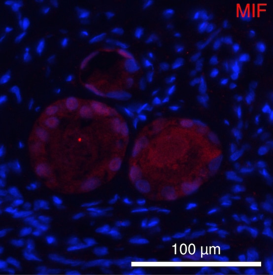 |
FH sarah (Verified Customer) (10-27-2023) | antibody worked great for WB
|
FH Sarah (Verified Customer) (01-24-2023) | This antibody worked well despite low protein concentration.
|
FH Emma (Verified Customer) (03-15-2022) | Nice antibody have used overnight @ 1:1000 on cell lysates and conditioned media. Produces a band at the correct size.
|
FH Ryan (Verified Customer) (02-27-2019) | Tissue was fixed in PFA with no additional antigen retrieval. Co-localisation with microglia based on known markers (not shown).
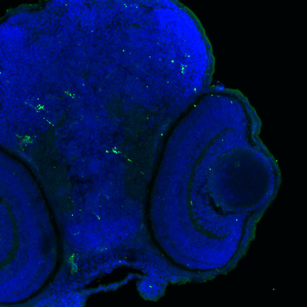 |

