Anticorps Polyclonal de lapin anti-MITF
MITF Polyclonal Antibody for WB, IHC, FC (Intra), IP, ELISA
Hôte / Isotype
Lapin / IgG
Réactivité testée
Humain, rat, souris
Applications
WB, IHC, IF, FC (Intra), IP, ELISA
Conjugaison
Non conjugué
N° de cat : 13092-1-AP
Synonymes
Galerie de données de validation
 at dilution of 1:1500 incubated at room temperature for 1.5 hours.")
 at dilution of 1:1000 incubated at room temperature for 1.5 hours.")
 at dilution of 1:1000 incubated at room temperature for 1.5 hours.")
 at dilution of 1:400 incubated at room temperature for 1.5 hours.")
 with mouse heart tissue lysate 4000ug.")
 at dilution of 1:1000 (under 10x lens). Heat mediated antigen retrieval with Tris-EDTA buffer (pH 9.0).")
 at dilution of 1:1000 (under 40x lens). Heat mediated antigen retrieval with Tris-EDTA buffer (pH 9.0).")
 and CoraLite®488-Conjugated Goat Anti-Rabbit IgG(H+L) (SA00013-2)(red), or 0.25 ug rabbit IgG isotype control (blue). Cells were fixed and permeabilized with Transcription Factor Staining Buffer Kit (PF00011).")
Applications testées
| Résultats positifs en WB | cellules A549, cellules Jurkat, cellules NIH/3T3, tissu cardiaque de souris, tissu cutané de rat |
| Résultats positifs en IP | tissu cardiaque de souris |
| Résultats positifs en IHC | tissu cutané de souris, il est suggéré de démasquer l'antigène avec un tampon de TE buffer pH 9.0; (*) À défaut, 'le démasquage de l'antigène peut être 'effectué avec un tampon citrate pH 6,0. |
| Résultats positifs en FC (Intra) | cellules HeLa, |
Dilution recommandée
| Application | Dilution |
|---|---|
| Western Blot (WB) | WB : 1:500-1:3000 |
| Immunoprécipitation (IP) | IP : 0.5-4.0 ug for 1.0-3.0 mg of total protein lysate |
| Immunohistochimie (IHC) | IHC : 1:500-1:2000 |
| Flow Cytometry (FC) (INTRA) | FC (INTRA) : 0.25 ug per 10^6 cells in a 100 µl suspension |
| It is recommended that this reagent should be titrated in each testing system to obtain optimal results. | |
| Sample-dependent, check data in validation data gallery | |
Applications publiées
| WB | See 32 publications below |
| IF | See 5 publications below |
Informations sur le produit
13092-1-AP cible MITF dans les applications de WB, IHC, IF, FC (Intra), IP, ELISA et montre une réactivité avec des échantillons Humain, rat, souris
| Réactivité | Humain, rat, souris |
| Réactivité citée | rat, Humain, souris |
| Hôte / Isotype | Lapin / IgG |
| Clonalité | Polyclonal |
| Type | Anticorps |
| Immunogène | MITF Protéine recombinante Ag3679 |
| Nom complet | microphthalmia-associated transcription factor |
| Masse moléculaire calculée | 91 aa, 10 kDa, 59 kDa |
| Poids moléculaire observé | 59-65 kDa |
| Numéro d’acquisition GenBank | BC012503 |
| Symbole du gène | MITF |
| Identification du gène (NCBI) | 4286 |
| Conjugaison | Non conjugué |
| Forme | Liquide |
| Méthode de purification | Purification par affinité contre l'antigène |
| Tampon de stockage | PBS with 0.02% sodium azide and 50% glycerol |
| Conditions de stockage | Stocker à -20°C. Stable pendant un an après l'expédition. L'aliquotage n'est pas nécessaire pour le stockage à -20oC Les 20ul contiennent 0,1% de BSA. |
Informations générales
The retinal pigment epithelium (RPE) has a essential role in maintaining visual function and dedifferentiation of RPE contributes to the pathophysiology of several ocular diseases[PMID: 22523078]. Microphthalmia-associated transcription factor (MITF) is a key regulator of RPE differentiation that is also down-regulated in dedifferentiated hfRPE cells. MITF is a basic helix-loop-helix (hHLH)-leucine zipper protein that involves in the development of various cell types, including neural crest-derived melanocytes and optic cup-derived retinal pigment epithelial cells [PMID: 10578055].
Protocole
| Product Specific Protocols | |
|---|---|
| WB protocol for MITF antibody 13092-1-AP | Download protocol |
| IHC protocol for MITF antibody 13092-1-AP | Download protocol |
| IP protocol for MITF antibody 13092-1-AP | Download protocol |
| Standard Protocols | |
|---|---|
| Click here to view our Standard Protocols |
Publications
| Species | Application | Title |
|---|---|---|
Cell Death Differ Lysine methylation of PPP1CA by the methyltransferase SUV39H2 disrupts TFEB-dependent autophagy and promotes intervertebral disc degeneration | ||
J Clin Invest mTORC1 feedback to AKT modulates lysosomal biogenesis through MiT/TFE regulation. | ||
Theranostics TFE3, a potential therapeutic target for Spinal Cord Injury via augmenting autophagy flux and alleviating ER stress. | ||
Phytomedicine Taxifolin inhibits melanoma proliferation/migration impeding USP18/Rac1/JNK/β-catenin oncogenic signaling | ||
Pigment Cell Melanoma Res D-tyrosine negatively regulates melanin synthesis by competitively inhibiting tyrosinase activity. |
Avis
The reviews below have been submitted by verified Proteintech customers who received an incentive for providing their feedback.
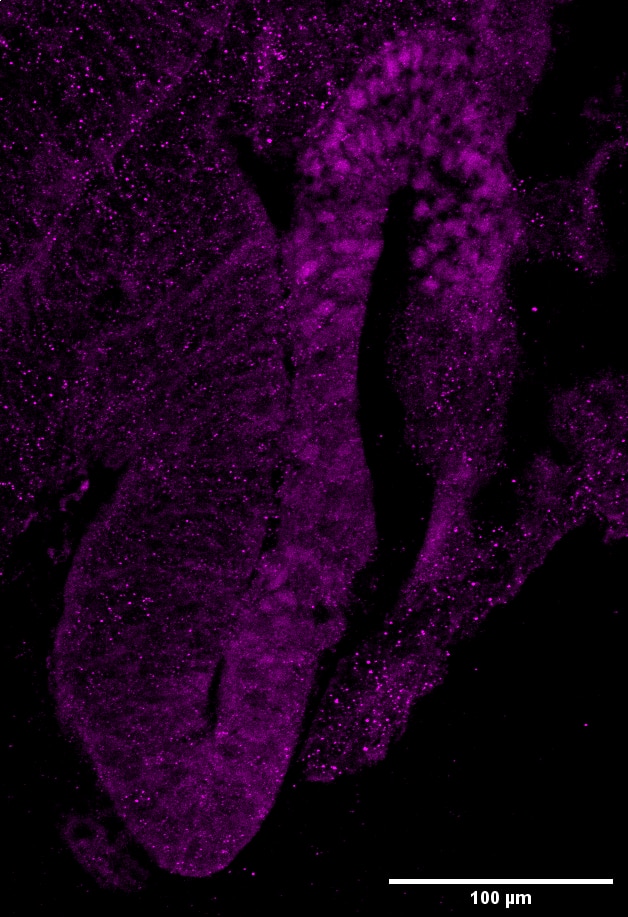
FH Eva (Verified Customer) (07-01-2025) | The antibody is functional both with and without antigen retrieval (AR: 110ºC for 1 min, a second cycle of 90ºC for 10s, in a citrate buffer 10mM pH=6); however, signal specificity appears reduced when antigen retrieval is applied, and its use is therefore not recommended. After x3 PBST (PBS + 0,2% Tween) washes, cryostat sample slides were incubated 2 hours with blocking buffer (PBST 0,2% + 5% FBS + 1% BSA) at Room Temperature (RT). The primary antibody was diluted 1:500 in Blocking Buffer and incubated with the samples overnight at 4 °C. Next day, wash x3 with PBST 0,2%, and incubate second antibody with fluorocrome at 1:500 2-3 hours at RT. Some background staining is observed, likely due to a high concentration of the secondary antibody, as it does not correspond to a specific signal. The staining is specifically localized in nuclei of pigmented cells, as expected, and no nonspecific signal is detected in other tissue types within the sample.
 |
FH Alessandro (Verified Customer) (11-06-2022) | no unspecific staining, great outcome
|

