- Phare
- Validé par KD/KO
Anticorps Monoclonal anti-MLKL
MLKL Monoclonal Antibody for WB, IHC, IF/ICC, IF-P, ELISA
Hôte / Isotype
Mouse / IgG1
Réactivité testée
Humain, souris et plus (2)
Applications
WB, IHC, IF/ICC, IF-P, IP, CoIP, ELISA
Conjugaison
Non conjugué
CloneNo.
3D4C6
N° de cat : 66675-1-Ig
Synonymes
Galerie de données de validation
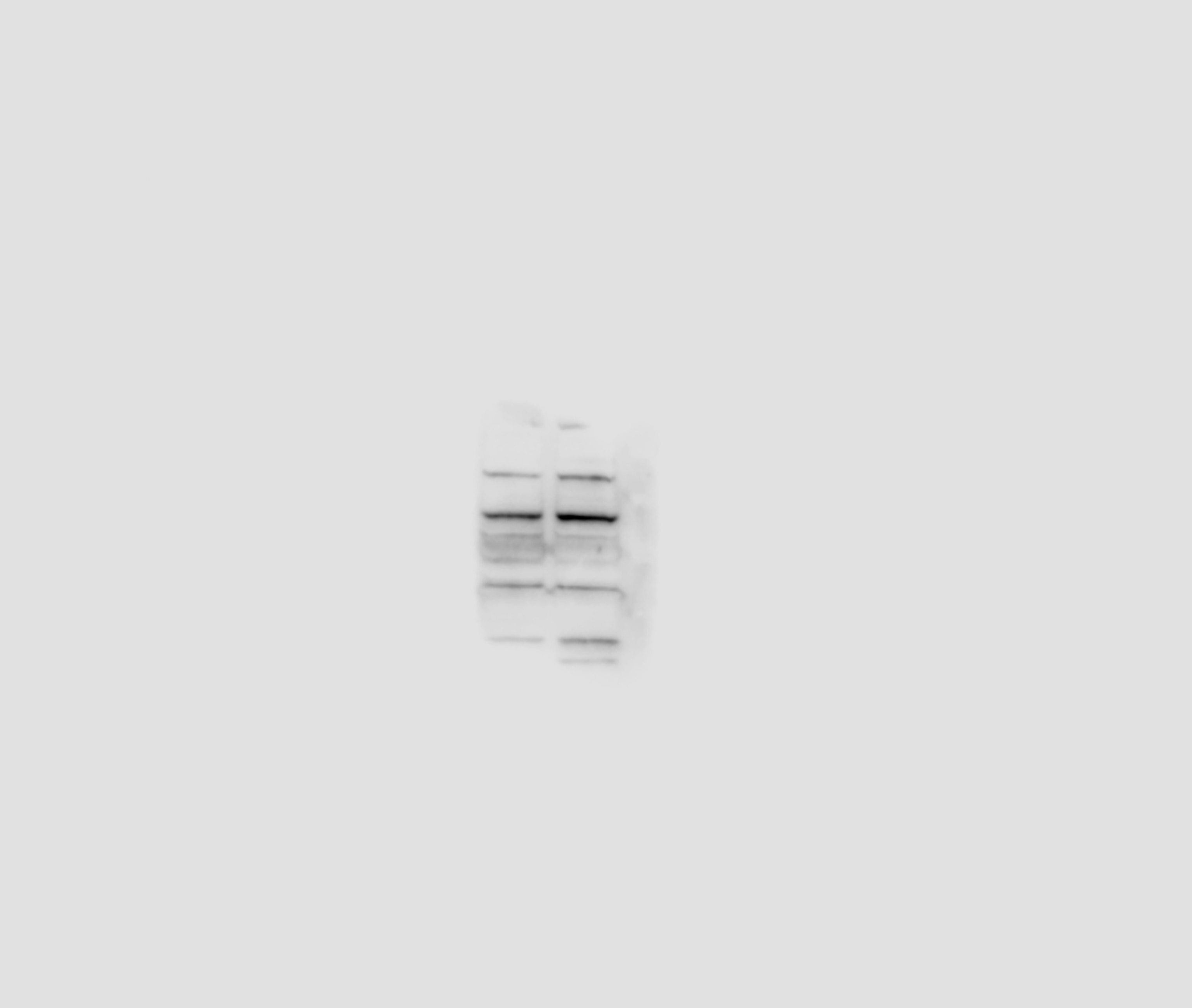
 at dilution of 1:10000 incubated at room temperature for 1.5 hours.")
 at dilution of 1:8000 incubated at room temperature for 1.5 hours.")
 at dilution of 1:3000 incubated at room temperature for 1.5 hours.")
 at dilution of 1:1000 (under 10x lens). Heat mediated antigen retrieval with Tris-EDTA buffer (pH 9.0).")
 at dilution of 1:1000 (under 40x lens). Heat mediated antigen retrieval with Tris-EDTA buffer (pH 9.0).")
 at dilution of 1:300 (under 10x lens. Heat mediated antigen retrieval with Tris-EDTA buffer (pH 9.0).")
 at dilution of 1:300 (under 40x lens. Heat mediated antigen retrieval with Tris-EDTA buffer (pH 9.0).")
 at dilution of 1:300 (under 10x lens. Heat mediated antigen retrieval with Tris-EDTA buffer (pH 9.0).")
 at dilution of 1:300 (under 40x lens. Heat mediated antigen retrieval with Tris-EDTA buffer (pH 9.0).")
 fixed human liver cancer tissue using 66675-1-Ig (MLKL antibody) at dilution of 1:100 and CoraLite488-Conjugated AffiniPure Goat Anti-Mouse IgG(H+L).")
 fixed HepG2 cells using MLKL antibody (66675-1-Ig, Clone: 3D4C6 ) at dilution of 1:800 and Multi-rAb CoraLite ® Plus 488-Goat Anti-Mouse Recombinant Secondary Antibody (H+L) (RGAM002).")
Applications testées
| Résultats positifs en WB | cellules HT-29, cellules 4T1, cellules HeLa, cellules HepG2, cellules NIH/3T3, cellules PC-3 |
| Résultats positifs en IHC | tissu de cancer du foie humain, tissu de cancer du côlon humain il est suggéré de démasquer l'antigène avec un tampon de TE buffer pH 9.0; (*) À défaut, 'le démasquage de l'antigène peut être 'effectué avec un tampon citrate pH 6,0. |
| Résultats positifs en IF-P | tissu de cancer du foie humain, |
| Résultats positifs en IF/ICC | cellules HepG2, |
Dilution recommandée
| Application | Dilution |
|---|---|
| Western Blot (WB) | WB : 1:5000-1:50000 |
| Immunohistochimie (IHC) | IHC : 1:500-1:2000 |
| Immunofluorescence (IF)-P | IF-P : 1:50-1:500 |
| Immunofluorescence (IF)/ICC | IF/ICC : 1:400-1:1600 |
| It is recommended that this reagent should be titrated in each testing system to obtain optimal results. | |
| Sample-dependent, check data in validation data gallery | |
Applications publiées
| KD/KO | See 4 publications below |
| WB | See 111 publications below |
| IHC | See 13 publications below |
| IF | See 18 publications below |
| IP | See 2 publications below |
| CoIP | See 2 publications below |
Informations sur le produit
66675-1-Ig cible MLKL dans les applications de WB, IHC, IF/ICC, IF-P, IP, CoIP, ELISA et montre une réactivité avec des échantillons Humain, souris
| Réactivité | Humain, souris |
| Réactivité citée | rat, Humain, poisson-zèbre, souris |
| Hôte / Isotype | Mouse / IgG1 |
| Clonalité | Monoclonal |
| Type | Anticorps |
| Immunogène | MLKL Protéine recombinante Ag15188 |
| Nom complet | mixed lineage kinase domain-like |
| Masse moléculaire calculée | 471 aa, 54 kDa |
| Poids moléculaire observé | 35-40 kDa, 50-55 kDa |
| Numéro d’acquisition GenBank | BC028141 |
| Symbole du gène | MLKL |
| Identification du gène (NCBI) | 197259 |
| Conjugaison | Non conjugué |
| Forme | Liquide |
| Méthode de purification | Purification par protéine G |
| Tampon de stockage | PBS with 0.02% sodium azide and 50% glycerol |
| Conditions de stockage | Stocker à -20°C. Stable pendant un an après l'expédition. L'aliquotage n'est pas nécessaire pour le stockage à -20oC Les 20ul contiennent 0,1% de BSA. |
Informations générales
Mixed lineage kinase domain like pseudokinase (MLKL), belongs to the protein kinase superfamily and has two MW of 54 and 30 kDa. MLKL plays a critical role in tumor necrosis factor (TNF)-induced necroptosis, a programmed cell death process, via interaction with receptor-interacting protein 3 (RIP3), which is a key signaling molecule in the necroptosis pathway. High levels of this protein and RIP3 are associated with inflammatory bowel disease in children. The 66675-1-Ig antibody recognizes 54 kDa MLKL monomer, 216 kDa MLKL tetramer, and a band around 45-50 kDa which are similar to papers published. (PMID: 31848291). MLKL can be detected as a 35kDa truncated isoform (PMID: 35965541).
Protocole
| Product Specific Protocols | |
|---|---|
| WB protocol for MLKL antibody 66675-1-Ig | Download protocol |
| IHC protocol for MLKL antibody 66675-1-Ig | Download protocol |
| IF protocol for MLKL antibody 66675-1-Ig | Download protocol |
| Standard Protocols | |
|---|---|
| Click here to view our Standard Protocols |
Publications
| Species | Application | Title |
|---|---|---|
Adv Sci (Weinh) Pressure Drives Rapid Burst-Like Coordinated Cellular Motion from 3D Cancer Aggregates. | ||
Acta Pharm Sin B Ligand-based substituent-anchoring design of selective receptor-interacting protein kinase 1 necroptosis inhibitors for ulcerative colitis therapy | ||
Acta Pharm Sin B A new perspective of triptolide-associated hepatotoxicity: the relevance of NF- κ B and NF- κ B-mediated cellular FLICE-inhibitory protein. | ||
Aging Dis MiR-29a-3p Improves Acute Lung Injury by Reducing Alveolar Epithelial Cell PANoptosis. | ||
Cell Death Dis S100A8/A9hi neutrophils induce mitochondrial dysfunction and PANoptosis in endothelial cells via mitochondrial complex I deficiency during sepsis | ||
Free Radic Biol Med Short-term exposure to low doses of aflatoxin B1 aggravates nonalcoholic steatohepatitis by TLR4-mediated necroptosis |
Avis
The reviews below have been submitted by verified Proteintech customers who received an incentive for providing their feedback.
FH Fan (Verified Customer) (03-22-2021) | Mouse retinal tissue was used. Multiple bands were detected. The target band has the strongest intensity.
 |
FH Paramananda (Verified Customer) (01-31-2019) | Excellent antibody!
|

