Anticorps Polyclonal de lapin anti-MYC tag
MYC tag Polyclonal Antibody for WB, IF/ICC, IP, ELISA
Hôte / Isotype
Lapin / IgG
Réactivité testée
Protéine recombinante et plus (4)
Applications
WB, IHC, IF/ICC, IP, CoIP, ChIP, RIP, ELISA
Conjugaison
Non conjugué
N° de cat : 16286-1-AP
Synonymes
 at dilution of 1:4000 incubated at room temperature for 1.5 hours.")
 with Transfected HEK-293 cells lysate 500 ug.")
 fixed Transfected HEK-293 cells using MYC tag antibody (16286-1-AP) at dilution of 1:400 and CoraLite®488-Conjugated AffiniPure Goat Anti-Rabbit IgG(H+L).")
 fixed Transfected HEK-293 cells using MYC tag antibody (16286-1-AP) at dilution of 1:800 and CoraLite®488-Conjugated AffiniPure Goat Anti-Rabbit IgG(H+L).")
.")
"MYC tag Antibodies" Comparison
View side-by-side comparison of MYC tag antibodies from other vendors to find the one that best suits your research needs.
Applications testées
| Résultats positifs en WB | protéine protéine recombinante, Protéine recombinante |
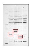
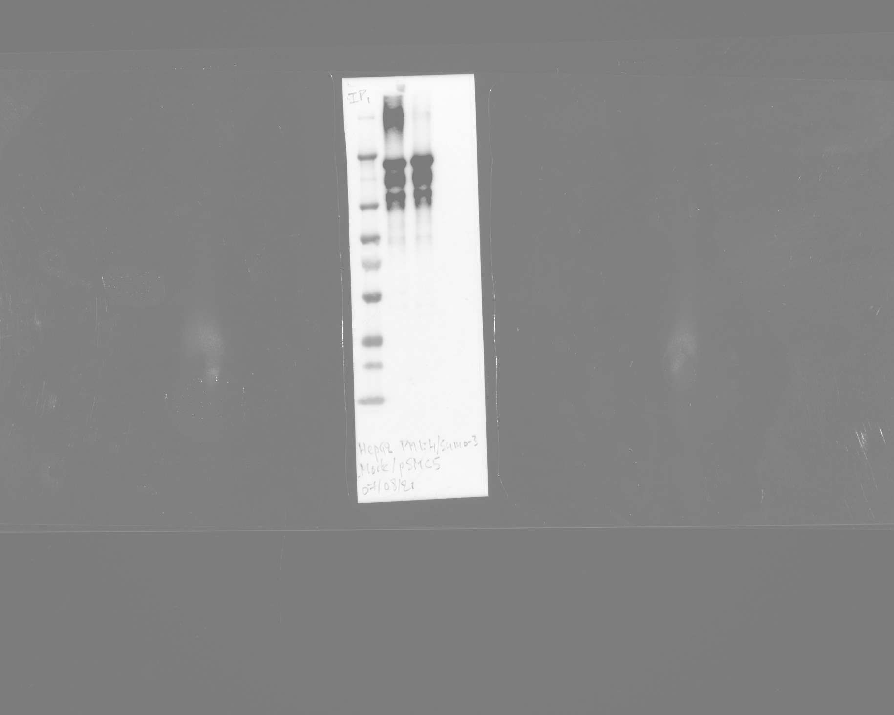
| Résultats positifs en IP | cellules HEK-293 transfectées, |
| Résultats positifs en IF/ICC | cellules HEK-293 transfectées, cellules MCF-7 |
Dilution recommandée
| Application | Dilution |
|---|---|
| Western Blot (WB) | WB : 1:1000-1:8000 |
| Immunoprécipitation (IP) | IP : 0.5-4.0 ug for 1.0-3.0 mg of total protein lysate |
| Immunofluorescence (IF)/ICC | IF/ICC : 1:200-1:800 |
| It is recommended that this reagent should be titrated in each testing system to obtain optimal results. | |
| Sample-dependent, check data in validation data gallery | |
Informations sur le produit
16286-1-AP cible MYC tag dans les applications de WB, IHC, IF/ICC, IP, CoIP, ChIP, RIP, ELISA et montre une réactivité avec des échantillons Protéine recombinante
| Réactivité | Protéine recombinante |
| Réactivité citée | Humain, porc, singe, souris |
| Hôte / Isotype | Lapin / IgG |
| Clonalité | Polyclonal |
| Type | Anticorps |
| Immunogène | MYC tag Protéine recombinante Ag9409 |
| Nom complet | Myc tag |
| Symbole du gène | Myc tag |
| Identification du gène (NCBI) | 99 |
| Conjugaison | Non conjugué |
| Forme | Liquide |
| Méthode de purification | Purification par affinité contre l'antigène |
| Tampon de stockage | PBS with 0.02% sodium azide and 50% glycerol |
| Conditions de stockage | Stocker à -20°C. Stable pendant un an après l'expédition. L'aliquotage n'est pas nécessaire pour le stockage à -20oC Les 20ul contiennent 0,1% de BSA. |
Informations générales
The myc-tag is a short synthetic polypeptide sequence derived from c-myc protein that can be added to recombinant proteins to enable isolation and study when an antibody is not available. This antibody recognizes the MYC tag EQKLISEEDL.
What is the molecular weight of myc?
1203Da: the ten amino acid myc tag sequence is EQKLISEEDL (in single letter code).
What are the applications for myc tag?
The addition of the myc tag to a particular protein can be useful when an antibody is not available to the protein of interest. Using recombinant DNA technology, the myc tag can be fused to the protein and then an antibody against the myc tag can be used to probe (PMID: 24490106). This is a reliable method and can be used in a number of different techniques, including purification using chromatography, tracking the protein in localization studies using immunofluorescence, or quantifying levels using Western Blot (PMID: 24490106).
What is the structure of myc tag?
The c-myc gene from which this tag is derived has a molecular weight of 49kDa, but the myc tag represents only a small portion of the C-terminus of this gene. The short polypeptide sequence can be fused to the N-terminus or the C-terminus of any protein without influencing function, although it is advised to avoid fusing it to a secretory signal. A cleavage site behind this tag is also sometimes engineered to allow removal with a specific protease.
Protocole
| Product Specific Protocols | |
|---|---|
| WB protocol for MYC tag antibody 16286-1-AP | Download protocol |
| IF protocol for MYC tag antibody 16286-1-AP | Download protocol |
| IP protocol for MYC tag antibody 16286-1-AP | Download protocol |
| Standard Protocols | |
|---|---|
| Click here to view our Standard Protocols |
Publications
| Species | Application | Title |
|---|---|---|
Signal Transduct Target Ther TRAF3 activates STING-mediated suppression of EV-A71 and target of viral evasion | ||
Signal Transduct Target Ther Circulating tumor cells shielded with extracellular vesicle-derived CD45 evade T cell attack to enable metastasis | ||
Nat Microbiol Ephrin receptor A2 is an epithelial cell receptor for Epstein-Barr virus entry. | ||
Drug Resist Updat MYC expression and fatty acid oxidation in EGFR-TKI acquired resistance | ||
Cell Stem Cell A primate-specific endogenous retroviral envelope protein sequesters SFRP2 to regulate human cardiomyocyte development | ||
Nat Commun TRIM5α recruits HDAC1 to p50 and Sp1 and promotes H3K9 deacetylation at the HIV-1 LTR |
Avis
The reviews below have been submitted by verified Proteintech customers who received an incentive for providing their feedback.
FH RASHMI (Verified Customer) (08-26-2024) | I used this antibody recently for ABC staining, it worked great, highly recommended.
|
FH RASHMI (Verified Customer) (07-26-2024) | used this product, highly recommended
|
FH Nazarine (Verified Customer) (02-01-2024) | Works well for Western Blot, IF, IP
|
FH Nazarine (Verified Customer) (02-01-2024) | Works well for Western Blot and IF and IP
|
FH Amy (Verified Customer) (03-05-2023) | Detected overexpressed Mac-tagged proteins in HEK293T cell lysates but with a fair few background bands.
 |
FH James (Verified Customer) (11-30-2022) | Very good antibody
 |
FH Boyan (Verified Customer) (08-03-2022) | gives a good signal in IF
|
FH Chahat (Verified Customer) (06-08-2022) | The myc antibody gives clear staining!
|
FH WEI (Verified Customer) (03-08-2022) | Good antibody for detecting overexpression proteins in 293 cells.
|
FH Minze (Verified Customer) (01-11-2022) | it works well for immunofluorecence so far.
|
FH Phoebe (Verified Customer) (03-01-2019) | This antibody was very clean in a western blot to detect Myc-tagged pol2 in S cerevisiae whole cell extract and chromatin fractionation samples.
|

