Anticorps Polyclonal de lapin anti-Myosin Light Chain 2/MLC-2V
Myosin Light Chain 2/MLC-2V Polyclonal Antibody for WB, IHC, IF-P, IF-Fro, FC (Intra), IP, ELISA
Hôte / Isotype
Lapin / IgG
Réactivité testée
Humain, poisson-zèbre, rat, souris et plus (2)
Applications
WB, IHC, IF-P, IF-Fro, FC (Intra), IP, ELISA
Conjugaison
Non conjugué
N° de cat : 10906-1-AP
Synonymes
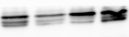
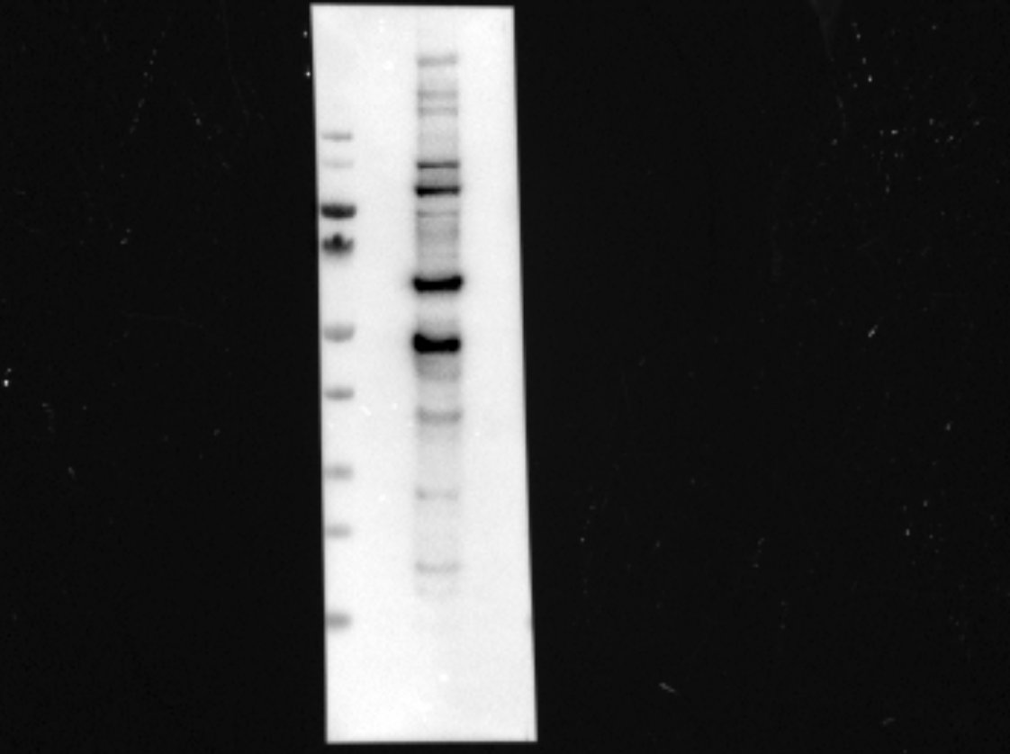
Galerie de données de validation
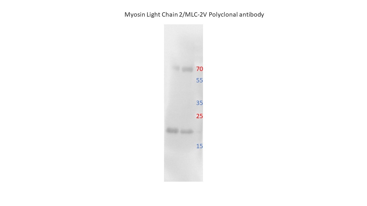
 at dilution of 1:8000 incubated at room temperature for 1.5 hours.")
 with mouse heart tissue lysate 6000ug.")
 at dilution of 1:800 (under 10x lens). Heat mediated antigen retrieval with Tris-EDTA buffer (pH 9.0).")
.")
 at dilution of 1:400 (under 40x lens). Heat mediated antigen retrieval with Tris-EDTA buffer (pH 9.0).")
 at dilution of 1:800 (under 40x lens). Heat mediated antigen retrieval with Tris-EDTA buffer (pH 9.0).")
.")
.")
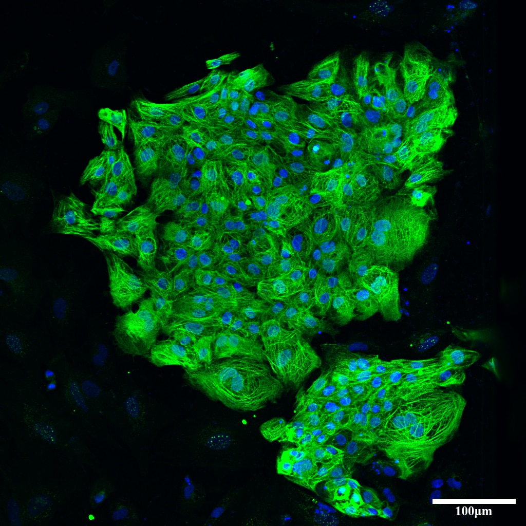
 fixed mouse heart tissue using Myosin Light Chain 2/MLC-2V antibody (10906-1-AP) at dilution of 1:200 and CoraLite®488-Conjugated AffiniPure Goat Anti-Rabbit IgG(H+L).")
 fixed paraffin-embedded mouse heart tissue using Myosin Light Chain 2/MLC-2V antibody (10906-1-AP) at dilution of 1:200 and Multi-rAb CoraLite ® Plus 488-Goat Anti-Rabbit Recombinant Secondary Antibody (H+L) (RGAR002). Heat mediated antigen retrieval with Tris-EDTA buffer (pH 9.0).")
 fixed frozen OCT-embedded mouse heart tissue using 10906-1-AP (Myosin Light Chain 2/MLC-2V antibody) at dilution of 1:200 and CoraLite488-Conjugated AffiniPure Goat Anti-Rabbit IgG(H+L).")
 and CoraLite®488-Conjugated AffiniPure Goat Anti-Rabbit IgG(H+L) at dilution 1:1000 (red), or 0.4 ug Control Antibody. Cells were fixed with 4% PFA and permeabilized with Flow Cytometry Perm Buffer (PF00011-C).")
Applications testées
| Résultats positifs en WB | tissu cardiaque de souris, tissu cardiaque de rat |
| Résultats positifs en IP | tissu cardiaque de souris |
| Résultats positifs en IHC | tissu cardiaque de souris, tissu cardiaque de rat, tissu cardiaque humain il est suggéré de démasquer l'antigène avec un tampon de TE buffer pH 9.0; (*) À défaut, 'le démasquage de l'antigène peut être 'effectué avec un tampon citrate pH 6,0. |
| Résultats positifs en IF-P | tissu cardiaque de souris, |
| Résultats positifs en IF-Fro | tissu cardiaque de souris, |
| Résultats positifs en FC (Intra) | cellules C2C12 |
Dilution recommandée
| Application | Dilution |
|---|---|
| Western Blot (WB) | WB : 1:2000-1:16000 |
| Immunoprécipitation (IP) | IP : 0.5-4.0 ug for 1.0-3.0 mg of total protein lysate |
| Immunohistochimie (IHC) | IHC : 1:400-1:1600 |
| Immunofluorescence (IF)-P | IF-P : 1:50-1:500 |
| Immunofluorescence (IF)-FRO | IF-FRO : 1:50-1:500 |
| Flow Cytometry (FC) (INTRA) | FC (INTRA) : 0.40 ug per 10^6 cells in a 100 µl suspension |
| It is recommended that this reagent should be titrated in each testing system to obtain optimal results. | |
| Sample-dependent, check data in validation data gallery | |
Applications publiées
| WB | See 66 publications below |
| IHC | See 11 publications below |
| IF | See 140 publications below |
| FC | See 2 publications below |
Informations sur le produit
10906-1-AP cible Myosin Light Chain 2/MLC-2V dans les applications de WB, IHC, IF-P, IF-Fro, FC (Intra), IP, ELISA et montre une réactivité avec des échantillons Humain, poisson-zèbre, rat, souris
| Réactivité | Humain, poisson-zèbre, rat, souris |
| Réactivité citée | rat, Humain, poisson-zèbre, porc, singe, souris |
| Hôte / Isotype | Lapin / IgG |
| Clonalité | Polyclonal |
| Type | Anticorps |
| Immunogène | Myosin Light Chain 2/MLC-2V Protéine recombinante Ag1356 |
| Nom complet | myosin, light chain 2, regulatory, cardiac, slow |
| Masse moléculaire calculée | 19 kDa |
| Poids moléculaire observé | 19 kDa |
| Numéro d’acquisition GenBank | BC015821 |
| Symbole du gène | Myosin Light Chain 2 |
| Identification du gène (NCBI) | 4633 |
| Conjugaison | Non conjugué |
| Forme | Liquide |
| Méthode de purification | Purification par affinité contre l'antigène |
| Tampon de stockage | PBS with 0.02% sodium azide and 50% glycerol |
| Conditions de stockage | Stocker à -20°C. Stable pendant un an après l'expédition. L'aliquotage n'est pas nécessaire pour le stockage à -20oC Les 20ul contiennent 0,1% de BSA. |
Informations générales
Background
MYL2 belongs to the myosin family of motor proteins. Their common features are ATP hydrolysis, actin binding, and potential for kinetic energy transduction. MYL2 stands for Myosin regulatory light chain 2, ventricular/cardiac muscle isoform (MLC-2), also known as the regulatory light chain of myosin (RLC). This isoform is distinct from those expressed in skeletal muscle (MYLPF), smooth muscle (MYL12B), and cardiac atrial muscle (MYL7) (PMID: 21345328). Due to its tissue specificity, MYL2 has been widely used as a marker of mature ventricular cardiomyocytes.
What is the molecular weight of MYL2?
There are two isoforms of this protein in humans that differ only slightly in length and produce proteins composed of 152 and 166 aa, which correspond to about 19 kDa (PMID: 16678204).
What is the subcellular localization of MYL2?
It is uniquely expressed in the cytoplasm of heart muscles. It colocalizes with actin cytoskeleton staining.
What is the tissue specificity of MYL2?
MYL2 is specifically expressed in ventricular cardiac tissue.
What is the function of MYL2 in cardiac tissue?
MYL2 is a contractile protein that plays a role in heart development and function. During cardiogenesis it plays an early role in cardiac contractility by promoting cardiac myofibril assembly and represents one of the earliest markers of ventricular specification (PMID 8506363).
In general, MYL2 interacts with the neck/tail region of the muscle thick filament protein myosin to regulate myosin motility and function (PMID 8316858). Its N-terminal domain is responsible for calcium and magnesium binding at their activating concentrations. This induces a functionally important conformational change (PMID: 18202317). However, the ion dissociation rate is not fast enough to modulate cardiac contractility on a beat-by-beat basis (PMIDs: 188447, 8804617). In addition, RLC function can be modulated by posttranslational modifications such as phosphorylation and deamidation in the N-terminal region. As a result, a significant change in the charge of the protein is introduced, which undoubtedly alters its interaction with the C-terminal myosin alpha-helical domain (PMID: 26074085).
What is MYL2's involvement in disease?
Diseases associated with MYL2 dysfunction include forms of familial hypertrophic cardiomyopathies, congenital fiber-type disproportion, and heart failure (PMID: 26074085).
Protocole
| Product Specific Protocols | |
|---|---|
| WB protocol for Myosin Light Chain 2/MLC-2V antibody 10906-1-AP | Download protocol |
| IHC protocol for Myosin Light Chain 2/MLC-2V antibody 10906-1-AP | Download protocol |
| IF protocol for Myosin Light Chain 2/MLC-2V antibody 10906-1-AP | Download protocol |
| IP protocol for Myosin Light Chain 2/MLC-2V antibody 10906-1-AP | Download protocol |
| Standard Protocols | |
|---|---|
| Click here to view our Standard Protocols |
Publications
| Species | Application | Title |
|---|---|---|
Science Conversion of human fibroblasts into functional cardiomyocytes by small molecules. | ||
Cell Stem Cell Cardiac repair in a porcine model of acute myocardial infarction with human induced pluripotent stem cell-derived cardiovascular cells. | ||
Cell Stem Cell Pathogenic LMNA variants disrupt cardiac lamina-chromatin interactions and de-repress alternative fate genes. | ||
Cell Stem Cell Gene editing to prevent ventricular arrhythmias associated with cardiomyocyte cell therapy | ||
Cell Stem Cell Gene editing to prevent ventricular arrhythmias associated with cardiomyocyte cell therapy | ||
Cell Stem Cell Long-term engraftment and maturation of autologous iPSC-derived cardiomyocytes in two rhesus macaques |
Avis
The reviews below have been submitted by verified Proteintech customers who received an incentive for providing their feedback.
FH Udesh (Verified Customer) (12-29-2023) | Worked well for WB at 1:1000 in Glioma cells
|
FH Brice-Emmanuel (Verified Customer) (12-18-2023) | Works very well in WB, even after more than 10 days at room temperature due to a delivery problem.
 |
FH Rebecca (Verified Customer) (10-28-2022) | 20ug of protein were loaded. Transference was performed at 4ºC and 90V for 2h. Antibody incubation was done ON at 4ºC.
 |
FH WEI (Verified Customer) (05-11-2022) | Strong, specific band for heart tissues
|
FH Irem (Verified Customer) (04-19-2022) | This antibody unspecifically recognizes at least four proteins in Jurkat cell lysate. The specific band, around 17 kDa, is so faint.
 |
FH Ning (Verified Customer) (02-20-2022) | This antibody works very well in immunofluorescence staining for iPSC-derived ventricular cardiomyocytes.
 |
FH Tongbin (Verified Customer) (08-25-2020) | This MLC2 antibody works very well in western blots.
|

