- Phare
- Validé par KD/KO
Anticorps Polyclonal de lapin anti-P21
P21 Polyclonal Antibody for WB, IHC, IF/ICC, IF-P, FC (Intra), IP, ELISA
Hôte / Isotype
Lapin / IgG
Réactivité testée
Humain et plus (4)
Applications
WB, IHC, IF/ICC, IF-P, FC (Intra), IP, CoIP, ELISA, Cell treatment
Conjugaison
Non conjugué
N° de cat : 10355-1-AP
Synonymes
Galerie de données de validation
 at dilution of 1:2000 incubated at room temperature for 1.5 hours.")
 at dilution of 1:3000 incubated at room temperature for 1.5 hours.")
 at dilution of 1:3000 incubated at room temperature for 1.5 hours.")
 at dilution of 1:800 incubated at room temperature for 1.5 hours.")
 at dilution of 1:600 incubated at room temperature for 1.5 hours.")
 at dilution of 1:2000 incubated at room temperature for 1.5 hours.")
 with MCF-7 cells lysate 1085 ug.")
 at dilution of 1:10000 (under 20x lens). Heat mediated antigen retrieval with Tris-EDTA buffer (pH 9.0).")
 at dilution of 1:10000 (under 20x lens). Heat mediated antigen retrieval with Tris-EDTA buffer (pH 9.0).")
 at dilution of 1:1000 (under 20x lens). Heat mediated antigen retrieval with Tris-EDTA buffer (pH 9.0).")
 at dilution of 1:1000 (under 20x lens). Heat mediated antigen retrieval with Tris-EDTA buffer (pH 9.0).")
 at dilution of 1:200 (under 10x lens. Heat mediated antigen retrieval with Tris-EDTA buffer (pH 9.0).")
 at dilution of 1:200 (under 40x lens. Heat mediated antigen retrieval with Tris-EDTA buffer (pH 9.0).")
 at dilution of 1:400 (under 10x lens. Heat mediated antigen retrieval with Tris-EDTA buffer (pH 9.0).")
 at dilution of 1:400 (under 40x lens. Heat mediated antigen retrieval with Tris-EDTA buffer (pH 9.0).")
 at dilution of 1:400 (under 10x lens. Heat mediated antigen retrieval with Tris-EDTA buffer (pH 9.0).")
 at dilution of 1:400 (under 40x lens. Heat mediated antigen retrieval with Tris-EDTA buffer (pH 9.0).")
 at dilution of 1:400 (under 10x lens. Heat mediated antigen retrieval with Tris-EDTA buffer (pH 9.0).")
 at dilution of 1:400 (under 40x lens. Heat mediated antigen retrieval with Tris-EDTA buffer (pH 9.0).")
 at dilution of 1:50 (under 10x lens).")
 at dilution of 1:50 (under 40x lens).")
 in the myotome of a somite at E11.5 by Dr. Zalc A. and Dr. Relaix F. (Red = p21; Green=Pax3 from a GFP reporter; Blue=DAPI).")
 fixed MCF-7 cells using P21 antibody (10355-1-AP) at dilution of 1:400 and CoraLite®594-Conjugated Goat Anti-Rabbit IgG(H+L) (SA00013-4), CL488-phalloidin (green).")
 at dilution of 1:50 and Alexa Fluor 488-conjugated AffiniPure Goat Anti-Rabbit IgG(H+L).")
 and CoraLite®488-Conjugated AffiniPure Goat Anti-Rabbit IgG(H+L) at dilution 1:1000 (red), or 0.4 ug Isotype Control. Cells were fixed and permeabilized with Transcription Factor Staining Buffer Kit (PF00011).")
Applications testées
| Résultats positifs en WB | cellules HUVEC, cellules HEK-293, cellules HeLa, cellules HepG2, cellules LNCaP, cellules MCF-7, cellules NCCIT, cellules THP-1, cellules U2OS |
| Résultats positifs en IP | cellules MCF-7, |
| Résultats positifs en IHC | tissu de cancer du côlon humain, tissu de cancer de la thyroïde humain, tissu de cancer de l'estomac humain, tissu de cancer du sein humain, tissu rénal humain il est suggéré de démasquer l'antigène avec un tampon de TE buffer pH 9.0; (*) À défaut, 'le démasquage de l'antigène peut être 'effectué avec un tampon citrate pH 6,0. |
| Résultats positifs en IF-P | tissu de myotome d'un somite à E11.5, |
| Résultats positifs en IF/ICC | cellules MCF-7, |
| Résultats positifs en FC (Intra) | cellules MCF-7 |
Dilution recommandée
| Application | Dilution |
|---|---|
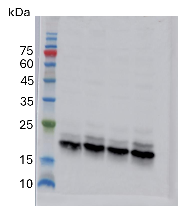
| Western Blot (WB) | WB : 1:1000-1:4000 |
| Immunoprécipitation (IP) | IP : 0.5-4.0 ug for 1.0-3.0 mg of total protein lysate |
| Immunohistochimie (IHC) | IHC : 1:5000-1:20000 |
| Immunofluorescence (IF)-P | IF-P : 1:10-1:100 |
| Immunofluorescence (IF)/ICC | IF/ICC : 1:200-1:800 |
| Flow Cytometry (FC) (INTRA) | FC (INTRA) : 0.40 ug per 10^6 cells in a 100 µl suspension |
| It is recommended that this reagent should be titrated in each testing system to obtain optimal results. | |
| Sample-dependent, check data in validation data gallery | |
Informations sur le produit
10355-1-AP cible P21 dans les applications de WB, IHC, IF/ICC, IF-P, FC (Intra), IP, CoIP, ELISA, Cell treatment et montre une réactivité avec des échantillons Humain
| Réactivité | Humain |
| Réactivité citée | bovin, canin, Humain, Lapin, porc |
| Hôte / Isotype | Lapin / IgG |
| Clonalité | Polyclonal |
| Type | Anticorps |
| Immunogène | P21 Protéine recombinante Ag0368 |
| Nom complet | cyclin-dependent kinase inhibitor 1A (p21, Cip1) |
| Masse moléculaire calculée | 18 kDa |
| Poids moléculaire observé | 21 kDa |
| Numéro d’acquisition GenBank | BC000275 |
| Symbole du gène | p21 |
| Identification du gène (NCBI) | 1026 |
| Conjugaison | Non conjugué |
| Forme | Liquide |
| Méthode de purification | Purification par affinité contre l'antigène |
| Tampon de stockage | PBS with 0.02% sodium azide and 50% glycerol |
| Conditions de stockage | Stocker à -20°C. Stable pendant un an après l'expédition. L'aliquotage n'est pas nécessaire pour le stockage à -20oC Les 20ul contiennent 0,1% de BSA. |
Informations générales
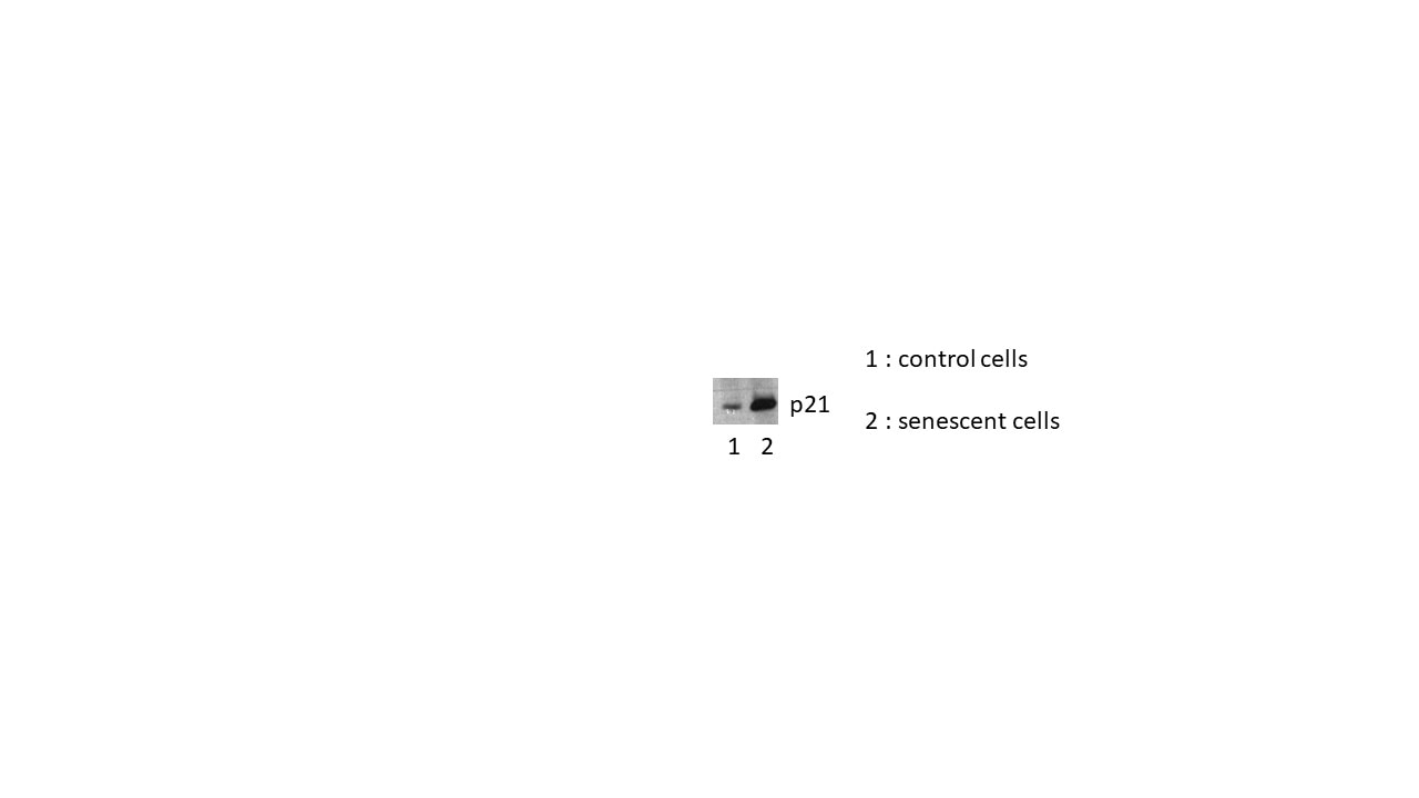
CDKN1A (p21, CIP1, WAF1) is a cyclin-dependent kinase inhibitor. CDKN1A binds to and inhibits the activity of cyclin-CDK2 or -CDK4 complexes, and thus functions as a regulator of cell cycle progression at the G1 phase. The expression of CDKN1A is induced by wild-type but not mutant p53 protein, through which CDKN1A mediates the p53-dependent cell cycle G1 phase arrest in response to a variety of stress stimuli. CDKN1A can interact with proliferating cell nuclear antigen (PCNA), and plays a regulatory role in S phase DNA replication and DNA damage repair. CDKN1A was reported to be specifically cleaved by CASP3-like caspases, which thus leads to a dramatic activation of CDK2, and may be instrumental in the execution of apoptosis following caspase activation. Two alternatively spliced variants, which encode an identical protein, have been reported.
Protocole
| Product Specific Protocols | |
|---|---|
| WB protocol for P21 antibody 10355-1-AP | Download protocol |
| IHC protocol for P21 antibody 10355-1-AP | Download protocol |
| IF protocol for P21 antibody 10355-1-AP | Download protocol |
| IP protocol for P21 antibody 10355-1-AP | Download protocol |
| Standard Protocols | |
|---|---|
| Click here to view our Standard Protocols |
Publications
| Species | Application | Title |
|---|---|---|
Gastroenterology Intestinal PPARα Protects Against Colon Carcinogenesis via Regulation of Methyltransferases DNMT1 and PRMT6. | ||
Sci Transl Med Guanosine diphosphate-mannose suppresses homologous recombination repair and potentiates antitumor immunity in triple-negative breast cancer | ||
Nat Commun Phosphoglycerate dehydrogenase activates PKM2 to phosphorylate histone H3T11 and attenuate cellular senescence | ||
Nat Commun URI alleviates tyrosine kinase inhibitors-induced ferroptosis by reprogramming lipid metabolism in p53 wild-type liver cancers | ||
Nat Commun Senescence-associated 13-HODE production promotes age-related liver steatosis by directly inhibiting catalase activity | ||
Mol Cancer CircVAPA promotes small cell lung cancer progression by modulating the miR-377-3p and miR-494-3p/IGF1R/AKT axis. |
Avis
The reviews below have been submitted by verified Proteintech customers who received an incentive for providing their feedback.
FH Ankush (Verified Customer) (08-13-2025) | The primary antibody was used to detect and quantify the expression level of the p21 protein in skin cells.
 |
FH Alexandros (Verified Customer) (06-06-2025) | Excellent performance; very nice staining on my membranes (PVDF) and not taking ages to develop as previous antibodies I had tried.
|
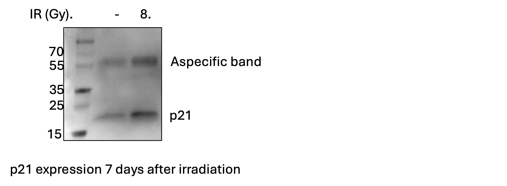
FH celine (Verified Customer) (11-19-2024) | I used in BSA 3% at 1:1000 for overnight incubation. It worked very well, but there is an unspecific band.
 |
FH Vikas (Verified Customer) (04-23-2024) | Used p21 antibody at 1:1000 dilution for western blotting worked great. Thank You. Highly recommended.
|
FH Hadil (Verified Customer) (05-24-2023) | I prepared this antibody in odyssey blocker at 1:1000 and incubated at 4 C overnight and seemed to get a good signal for p21. There were some non-specific bands properly caused by long incubation periods. So, i would recommend using it at this concentration for 1.5-2 hr at RT.
|
FH Malak (Verified Customer) (04-22-2021) | The best incubation period for this antibody is 1.5 hr at room temperature, longer incubations (especially overnight at 4°C) may result in aspecific bands.
|
FH Susan (Verified Customer) (12-03-2019) | We had been having issues with our old P21 antibody and this one worked great.
|
FH Béatrice (Verified Customer) (09-04-2019) | This is an excellent and specific antibody, without background.When prepared in 3%BSA, it can be reused several times and kept at-20°C
 |

