- Phare
- Validé par KD/KO
Anticorps Polyclonal de lapin anti-PABPC1,PABP
PABPC1,PABP Polyclonal Antibody for WB, IHC, IF/ICC, IP, ELISA
Hôte / Isotype
Lapin / IgG
Réactivité testée
Humain, rat, souris et plus (1)
Applications
WB, IHC, IF/ICC, IP, CoIP, RIP, ELISA
Conjugaison
Non conjugué
N° de cat : 10970-1-AP
Synonymes
Galerie de données de validation
 at dilution of 1:2500 incubated at room temperature for 1.5 hours.")
 at dilution of 1:6000 incubated at room temperature for 1.5 hours.")
 at dilution of 1:8000 incubated at room temperature for 1.5 hours.")
 at dilution of 1:6000 incubated at room temperature for 1.5 hours.")
 at dilution of 1:1000 incubated at room temperature for 1.5 hours.")
 at dilution of 1:500 incubated at room temperature for 1.5 hours.")
 at dilution of 1:200 incubated at room temperature for 1.5 hours.")
 at dilution of 1:300 incubated at room temperature for 1.5 hours.")
 at dilution of 1:300 incubated at room temperature for 1.5 hours.")
 at dilution of 1:200 incubated at room temperature for 1.5 hours.")
 with mouse testis tissue lysate 4000ug.")
 at dilution of 1:50 (under 40x lens).")
 at dilution of 1:50.")
 at dilution of 1:50.")
 at dilution of 1:50 (under 10x lens).")
 fixed MCF-7 cells using PABPC1,PABP antibody (10970-1-AP) at dilution of 1:400 and CoraLite®488-Conjugated Goat Anti-Rabbit IgG(H+L) (SA00013-2).")
Applications testées
| Résultats positifs en WB | cellules HCT 116, cellules A549, cellules HeLa, cellules HT-29, cellules MCF-7, cellules NCI-H1299, cellules PC-3, tissu testiculaire de rat, tissu testiculaire de souris |
| Résultats positifs en IP | tissu testiculaire de souris |
| Résultats positifs en IHC | tissu pancréatique humain, tissu testiculaire humain il est suggéré de démasquer l'antigène avec un tampon de TE buffer pH 9.0; (*) À défaut, 'le démasquage de l'antigène peut être 'effectué avec un tampon citrate pH 6,0. |
| Résultats positifs en IF/ICC | cellules MCF-7, |
Dilution recommandée
| Application | Dilution |
|---|---|
| Western Blot (WB) | WB : 1:1000-1:5000 |
| Immunoprécipitation (IP) | IP : 0.5-4.0 ug for 1.0-3.0 mg of total protein lysate |
| Immunohistochimie (IHC) | IHC : 1:20-1:200 |
| Immunofluorescence (IF)/ICC | IF/ICC : 1:200-1:800 |
| It is recommended that this reagent should be titrated in each testing system to obtain optimal results. | |
| Sample-dependent, check data in validation data gallery | |
Informations sur le produit
10970-1-AP cible PABPC1,PABP dans les applications de WB, IHC, IF/ICC, IP, CoIP, RIP, ELISA et montre une réactivité avec des échantillons Humain, rat, souris
| Réactivité | Humain, rat, souris |
| Réactivité citée | rat, Humain, porc, souris |
| Hôte / Isotype | Lapin / IgG |
| Clonalité | Polyclonal |
| Type | Anticorps |
| Immunogène | PABPC1,PABP Protéine recombinante Ag1422 |
| Nom complet | poly(A) binding protein, cytoplasmic 1 |
| Masse moléculaire calculée | 71 kDa |
| Poids moléculaire observé | 71 kDa |
| Numéro d’acquisition GenBank | BC015958 |
| Symbole du gène | PABPC1 |
| Identification du gène (NCBI) | 26986 |
| Conjugaison | Non conjugué |
| Forme | Liquide |
| Méthode de purification | Purification par affinité contre l'antigène |
| Tampon de stockage | PBS with 0.02% sodium azide and 50% glycerol |
| Conditions de stockage | Stocker à -20°C. Stable pendant un an après l'expédition. L'aliquotage n'est pas nécessaire pour le stockage à -20oC Les 20ul contiennent 0,1% de BSA. |
Informations générales
The poly(A)-binding protein (PABP), which is found complexed to the 3-prime poly(A) tail of eukaryotic mRNA, is required for poly(A) shortening and translation initiation [PMID: 21989405]. Polyadenylate-binding protein 1 (PABPC1) is a cytoplasmic-nuclear shuttling protein important for protein translation initiation, and both RNA processing and stability. In the cytoplasm, PABPC1 binds to the 3' poly(A) tail of eukaryotic mRNAs through its RNA-recognition motifs (RRM) and interacts with the N-terminus of eIF4G, part of the eIF4F complex associated with the 5' cap structure [PMID:20009508, 17381337].
Protocole
| Product Specific Protocols | |
|---|---|
| WB protocol for PABPC1,PABP antibody 10970-1-AP | Download protocol |
| IHC protocol for PABPC1,PABP antibody 10970-1-AP | Download protocol |
| IF protocol for PABPC1,PABP antibody 10970-1-AP | Download protocol |
| IP protocol for PABPC1,PABP antibody 10970-1-AP | Download protocol |
| Standard Protocols | |
|---|---|
| Click here to view our Standard Protocols |
Publications
| Species | Application | Title |
|---|---|---|
Brain Behav Immun Transcriptomic and proteomic profiling of bi-partite and tri-partite murine iPSC-derived neurospheroids under steady-state and inflammatory condition | ||
Adv Sci (Weinh) Primate-Specific DAZ Regulates Translation of Cell Proliferation-Related mRNAs and is Essential for Maintenance of Spermatogonia | ||
Exp Mol Med The deubiquitinating enzyme STAMBP is a newly discovered driver of triple-negative breast cancer progression that maintains RAI14 protein stability | ||
Nat Commun An oncopeptide regulates m6A recognition by the m6A reader IGF2BP1 and tumorigenesis. | ||
Dev Cell DDX20 is required for cell-cycle reentry of prospermatogonia and establishment of spermatogonial stem cell pool during testicular development in mice | ||
J Exp Med E3 ligase MKRN3 is a tumor suppressor regulating PABPC1 ubiquitination in non-small cell lung cancer. |
Avis
The reviews below have been submitted by verified Proteintech customers who received an incentive for providing their feedback.
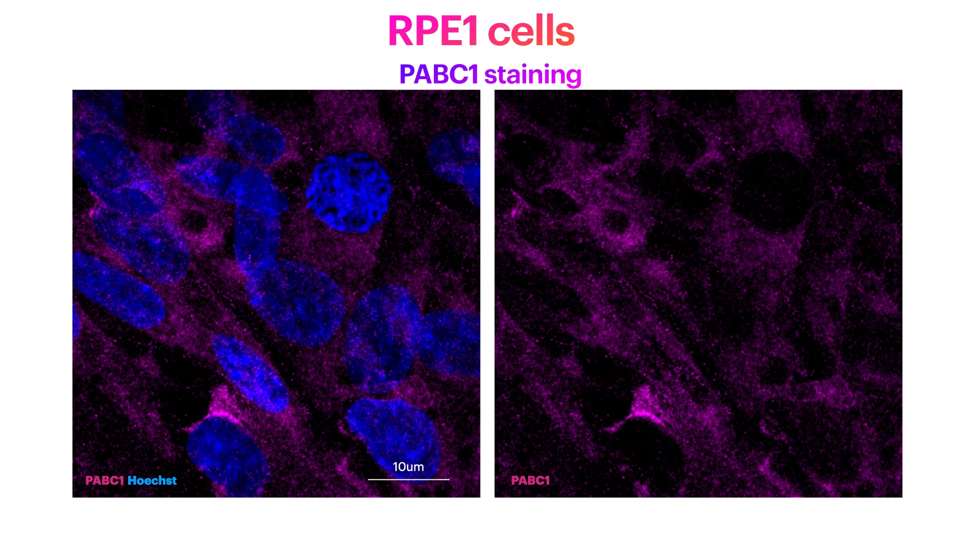
FH Elisa (Verified Customer) (01-13-2023) | PABPC1 (Poly(A) Binding Protein Cytoplasmic 1) staining (in magenta) and Hoechst (nuclear staining) in blue. PABPC1 ab shows a clear citoplasma staining. Method: RPE1 cells were fixed in cold methanol for 10' at -20C. Cells were then rehydrated with PBS for 5'. Membrane permeabilization was then performed with 0.1% Triton + 0.1% Tween +0.01%SDS in PBS for 10'. Cells were finally incubated with blocking buffer (5% BSA+ 0.1% Tween in PBS) for 30' at RT. Primary antibody was diluted in blocking buffer 1:200 and incubated for 1h at room temperature. Alexa-488-Anti-rabbit was used as secondary antibody (1:600 dilution) (1h at room temperature).
 |
FH Zee (Verified Customer) (01-28-2020) | It worked very well when I performed western blot.
|
FH George (Verified Customer) (09-11-2019) | Rb PABP Ig shows very little endogenous background staining with clear dispersed staining seen both in the nucleus and cytoplasm. 90mins heat shock at 42oC led to granular formation in the cytoplasm
|

