- Phare
- Validé par KD/KO
Anticorps Polyclonal de lapin anti-PARD3
PARD3 Polyclonal Antibody for WB, IHC, IF/ICC, IF-P, IP, ELISA
Hôte / Isotype
Lapin / IgG
Réactivité testée
Humain, souris et plus (2)
Applications
WB, IHC, IF/ICC, IF-P, IP, CoIP, ELISA
Conjugaison
Non conjugué
N° de cat : 11085-1-AP
Synonymes
Galerie de données de validation
 at dilution of 1:5000 incubated at room temperature for 1.5 hours.")
 at dilution of 1:300 incubated at room temperature for 1.5 hours.")
 with MCF-7 cells lysate 2900ug.")
 at dilution of 1:50 (under 10x lens).")
 at dilution of 1:50 (under 40x lens).")
 fixed mouse kidney tissue using 11085-1-AP (PARD3 antibody) at dilution of 1:50 and Alexa Fluor 488-conjugated Goat Anti-Rabbit IgG(H+L).")
 fixed mouse brain tissue using 11085-1-AP (PARD3 antibody) at dilution of 1:50 and Alexa Fluor 488-conjugated Goat Anti-Rabbit IgG(H+L).")
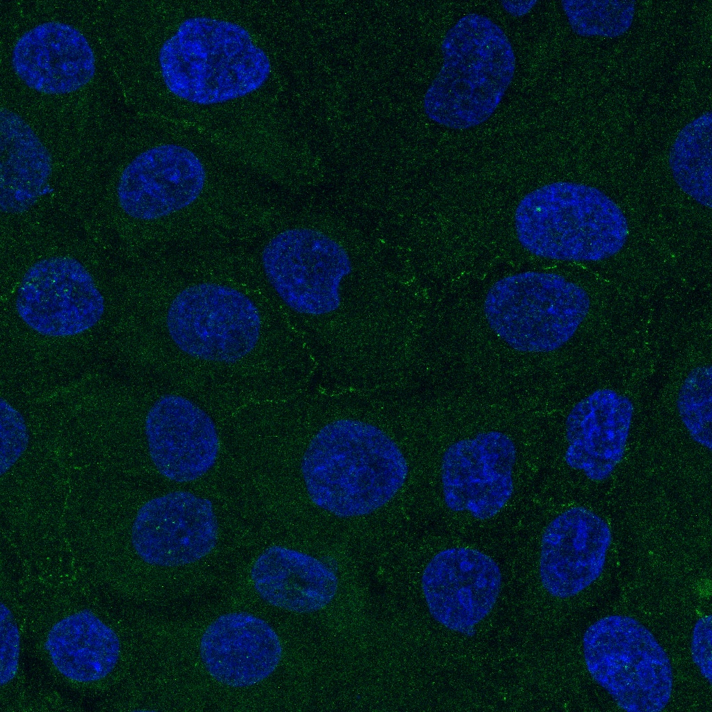
 fixed MCF-7 cells using PARD3 antibody (11085-1-AP) at dilution of 1:400 and CoraLite®488-Conjugated Goat Anti-Rabbit IgG(H+L).")
Applications testées
| Résultats positifs en WB | cellules A549, cellules HEK-293, cellules HeLa, cellules MCF-7 |
| Résultats positifs en IP | cellules MCF-7 |
| Résultats positifs en IHC | tissu de cancer du côlon humain, il est suggéré de démasquer l'antigène avec un tampon de TE buffer pH 9.0; (*) À défaut, 'le démasquage de l'antigène peut être 'effectué avec un tampon citrate pH 6,0. |
| Résultats positifs en IF-P | tissu rénal de souris, tissu cérébral de souris |
| Résultats positifs en IF/ICC | cellules MCF-7, |
Dilution recommandée
| Application | Dilution |
|---|---|
| Western Blot (WB) | WB : 1:2000-1:10000 |
| Immunoprécipitation (IP) | IP : 0.5-4.0 ug for 1.0-3.0 mg of total protein lysate |
| Immunohistochimie (IHC) | IHC : 1:50-1:200 |
| Immunofluorescence (IF)-P | IF-P : 1:50-1:500 |
| Immunofluorescence (IF)/ICC | IF/ICC : 1:200-1:800 |
| It is recommended that this reagent should be titrated in each testing system to obtain optimal results. | |
| Sample-dependent, check data in validation data gallery | |
Applications publiées
| KD/KO | See 4 publications below |
| WB | See 13 publications below |
| IHC | See 6 publications below |
| IF | See 12 publications below |
| IP | See 2 publications below |
| CoIP | See 1 publications below |
Informations sur le produit
11085-1-AP cible PARD3 dans les applications de WB, IHC, IF/ICC, IF-P, IP, CoIP, ELISA et montre une réactivité avec des échantillons Humain, souris
| Réactivité | Humain, souris |
| Réactivité citée | rat, Humain, poulet, souris |
| Hôte / Isotype | Lapin / IgG |
| Clonalité | Polyclonal |
| Type | Anticorps |
| Immunogène | PARD3 Protéine recombinante Ag1565 |
| Nom complet | par-3 partitioning defective 3 homolog (C. elegans) |
| Masse moléculaire calculée | 151 kDa |
| Poids moléculaire observé | 180 kDa, 140-150 kDa, 100 kDa |
| Numéro d’acquisition GenBank | BC011711 |
| Symbole du gène | PARD3 |
| Identification du gène (NCBI) | 56288 |
| Conjugaison | Non conjugué |
| Forme | Liquide |
| Méthode de purification | Purification par affinité contre l'antigène |
| Tampon de stockage | PBS with 0.02% sodium azide and 50% glycerol |
| Conditions de stockage | Stocker à -20°C. Stable pendant un an après l'expédition. L'aliquotage n'est pas nécessaire pour le stockage à -20oC Les 20ul contiennent 0,1% de BSA. |
Informations générales
PARD3 (also known as ASIP, Par3, or Bazooka) is one of PARD proteins which are essential for asymmetric cell division and polarized growth. PARD3 is involved in the establishment of cell polarity and in the asymmetric cytokinesis. It plays a role in tight junctions at epithelial cell-cell contacts. PARD3 has three splice isoforms at 100 kDa, 150 kDa, and 180 kDa. This polyclonal antibody raised against C-terminal 281 amino acids of human PARD3 recognizes these three isoforms.
Protocole
| Product Specific Protocols | |
|---|---|
| WB protocol for PARD3 antibody 11085-1-AP | Download protocol |
| IHC protocol for PARD3 antibody 11085-1-AP | Download protocol |
| IF protocol for PARD3 antibody 11085-1-AP | Download protocol |
| IP protocol for PARD3 antibody 11085-1-AP | Download protocol |
| Standard Protocols | |
|---|---|
| Click here to view our Standard Protocols |
Publications
| Species | Application | Title |
|---|---|---|
Adv Mater m6 A Reader YTHDF1-Targeting Engineered Small Extracellular Vesicles for Gastric Cancer Therapy via Epigenetic and Immune Regulation | ||
Matrix Biol Ameloblastin promotes polarization of ameloblast cell lines in a 3-D cell culture system | ||
Development Coupling of apical-basal polarity and PCP to interpret the Wnt signaling gradient and orient feather branch.
| ||
FASEB J Perturbation of epithelial apicobasal polarity by rhomboid family-1 gene overexpression. | ||
iScience Glucocorticoid receptor-mediated Nr1d1 chromatin circadian misalignment in stress-induced irritable bowel syndrome |
Avis
The reviews below have been submitted by verified Proteintech customers who received an incentive for providing their feedback.
FH Sarah (Verified Customer) (06-26-2019) | Western blot: Total cell lysate (15 ug) was resolved on a 4-12% Bis-Tris gel and transferred to nitrocellulose membrane. Membrane was incubated in blocking buffer (5% milk/0.1% Tween-20) for 1h. Membrane was incubated with anti-PARD-3 in blocking buffer (1:1000) at 4C overnight. After washing, membrane was incubated in anti-rabbit-HRP in blocking bufffer (1:3000) for 1h at room temperature. Protein was detected using ECL reagent and imaged on a chemiluminescence detection system.Immunofluorescence: Cells fixed in MeOH at -20 degrees were stained with 1:200 primary antibody. After washing, coverslips were incubated in 1:500 AF488 secondary antibody. Following mounting, images were acquired using a confocal microscope. Staining was very weak, as is seen by the image (Green = PARD3, Blue = Hoescht)
 |

