- Phare
- Validé par KD/KO
Anticorps Polyclonal de lapin anti-PCGF6
PCGF6 Polyclonal Antibody for WB, IHC, FC (Intra), IP, ELISA
Hôte / Isotype
Lapin / IgG
Réactivité testée
Humain, souris
Applications
WB, IHC, FC (Intra), IP, CoIP, ChIP, ELISA
Conjugaison
Non conjugué
N° de cat : 24103-1-AP
Synonymes
Galerie de données de validation
 at dilution of 1:1000 incubated at room temperature for 1.5 hours.")
 with sh-Control and sh-PCGF6 transfected HEK-293 cells.")
 with HEK-293T cells lysate 2200 ug.")
 at dilution of 1:300 (under 40x lens). Heat mediated antigen retrieval with Tris-EDTA buffer (pH 9.0).")
 at dilution of 1:300 (under 10x lens). Heat mediated antigen retrieval with Tris-EDTA buffer (pH 9.0).")
 and CoraLite®488-Conjugated AffiniPure Goat Anti-Rabbit IgG(H+L) at dilution 1:1000 (red), or 0.4 ug Isotype Control. Cells were fixed and permeabilized with Transcription Factor Staining Buffer Kit (PF00011).")
Applications testées
| Résultats positifs en WB | cellules HEK-293, cellules HEK-293T, cellules PC-13, tissu hépatique de souris |
| Résultats positifs en IP | cellules HEK-293T, |
| Résultats positifs en IHC | tissu testiculaire de souris, il est suggéré de démasquer l'antigène avec un tampon de TE buffer pH 9.0; (*) À défaut, 'le démasquage de l'antigène peut être 'effectué avec un tampon citrate pH 6,0. |
| Résultats positifs en FC (Intra) | cellules HEK-293T |
Dilution recommandée
| Application | Dilution |
|---|---|
| Western Blot (WB) | WB : 1:1000-1:4800 |
| Immunoprécipitation (IP) | IP : 0.5-4.0 ug for 1.0-3.0 mg of total protein lysate |
| Immunohistochimie (IHC) | IHC : 1:150-1:600 |
| Flow Cytometry (FC) (INTRA) | FC (INTRA) : 0.40 ug per 10^6 cells in a 100 µl suspension |
| It is recommended that this reagent should be titrated in each testing system to obtain optimal results. | |
| Sample-dependent, check data in validation data gallery | |
Applications publiées
| KD/KO | See 3 publications below |
| WB | See 8 publications below |
| IP | See 2 publications below |
| CoIP | See 1 publications below |
| ChIP | See 2 publications below |
Informations sur le produit
24103-1-AP cible PCGF6 dans les applications de WB, IHC, FC (Intra), IP, CoIP, ChIP, ELISA et montre une réactivité avec des échantillons Humain, souris
| Réactivité | Humain, souris |
| Réactivité citée | Humain, souris |
| Hôte / Isotype | Lapin / IgG |
| Clonalité | Polyclonal |
| Type | Anticorps |
| Immunogène | PCGF6 Protéine recombinante Ag21124 |
| Nom complet | polycomb group ring finger 6 |
| Masse moléculaire calculée | 352 aa, 39 kDa |
| Poids moléculaire observé | 40-50 kDa |
| Numéro d’acquisition GenBank | BC010235 |
| Symbole du gène | PCGF6 |
| Identification du gène (NCBI) | 84108 |
| Conjugaison | Non conjugué |
| Forme | Liquide |
| Méthode de purification | Purifié par affinité contre l'antigène |
| Tampon de stockage | PBS with 0.02% sodium azide and 50% glycerol |
| Conditions de stockage | Stocker à -20°C. Stable pendant un an après l'expédition. L'aliquotage n'est pas nécessaire pour le stockage à -20oC Les 20ul contiennent 0,1% de BSA. |
Informations générales
PCGF6, also named as MBLR or RNF134, is a 350 amino acid protein, which contains one RING-type zinc finger. PCGF6 localizes in the nucleus and is widely expressed in many tissues. PCGF6 as a transcriptional repressor may modulate the levels of histone H3K4Me3 by activating KDM5D histone demethylase.
Protocole
| Product Specific Protocols | |
|---|---|
| WB protocol for PCGF6 antibody 24103-1-AP | Download protocol |
| IHC protocol for PCGF6 antibody 24103-1-AP | Download protocol |
| IP protocol for PCGF6 antibody 24103-1-AP | Download protocol |
| Standard Protocols | |
|---|---|
| Click here to view our Standard Protocols |
Publications
| Species | Application | Title |
|---|---|---|
Cell Stem Cell SUMO Safeguards Somatic and Pluripotent Cell Identities by Enforcing Distinct Chromatin States. | ||
Sci Adv The SAM domain-containing protein 1 (SAMD1) acts as a repressive chromatin regulator at unmethylated CpG islands. | ||
Nat Commun E2F6 initiates stable epigenetic silencing of germline genes during embryonic development. | ||
Nat Commun PCGF6 controls neuroectoderm specification of human pluripotent stem cells by activating SOX2 expression.
| ||
Nat Commun Repression of germline genes by PRC1.6 and SETDB1 in the early embryo precedes DNA methylation-mediated silencing.
| ||
Elife Loss of MGA repression mediated by an atypical polycomb complex promotes tumor progression and invasiveness. |
Avis
The reviews below have been submitted by verified Proteintech customers who received an incentive for providing their feedback.
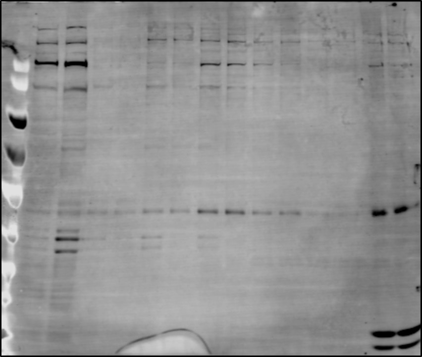
FH Aktan (Verified Customer) (12-24-2019) | The blot is subcellular fractionation of the cell nuclei using increasing concentrations of salt. Last two lanes are chromatin fractions. After electrophoresis and transfer, 5%BSA in PBST was used as blocker for 1h. The primary antibody is diluted in blocker 1:500 and the blot is incubated o/n at cold. The bands are detected using licor secondary antibodies and Licor imager.
 |

