- Phare
- Validé par KD/KO
Anticorps Monoclonal anti-PCNA
PCNA Monoclonal Antibody for WB, IHC, IF/ICC, IP, ELISA
Hôte / Isotype
Mouse / IgG1
Réactivité testée
Humain, porc, rat, souris et plus (2)
Applications
WB, IHC, IF/ICC, IP, CoIP, ELISA
Conjugaison
Non conjugué
CloneNo.
10D10E11
N° de cat : 60097-1-Ig
Synonymes
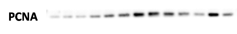
 incubated at room temperature for 1.5 hours.")
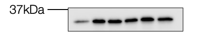
 at various dilution incubated at room temperature for 1.5 hours.")
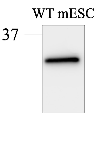
 with sh-Control and sh-PCNA transfected HEK-293 cells.")
 at dilution of 1:1000 incubated at room temperature for 1.5 hours.")
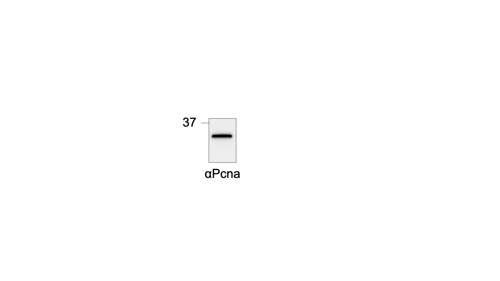
 with HepG2 cells lysate 3000ug.")
 at dilution of 1:1000 (under 10x lens. Heat mediated antigen retrieval with Tris-EDTA buffer (pH 9.0).")
 at dilution of 1:2000 (under 10x lens). Heat mediated antigen retrieval with Tris-EDTA buffer (pH 9.0).")
 at dilution of 1:16000 (under 40x lens). Heat mediated antigen retrieval with Tris-EDTA buffer (pH 9.0).")
 at dilution of 1:1000 (under 40x lens. Heat mediated antigen retrieval with Tris-EDTA buffer (pH 9.0).")
 at dilution of 1:1000 (under 10x lens. Heat mediated antigen retrieval with Tris-EDTA buffer (pH 9.0).")
 at dilution of 1:1000 (under 40x lens. Heat mediated antigen retrieval with Tris-EDTA buffer (pH 9.0).")
 at dilution of 1:800 (under 10x lens).")
 at dilution of 1:800 (under 40x lens).")
 at dilution of 1:2000 (under 10x lens). Heat mediated antigen retrieval with Tris-EDTA buffer (pH 9.0).")
 at dilution of 1:2000 (under 40x lens). Heat mediated antigen retrieval with Tris-EDTA buffer (pH 9.0).")
 at dilution of 1:2000 (under 10x lens). Heat mediated antigen retrieval with Tris-EDTA buffer (pH 9.0).")
 at dilution of 1:2000 (under 40x lens). Heat mediated antigen retrieval with Tris-EDTA buffer (pH 9.0).")
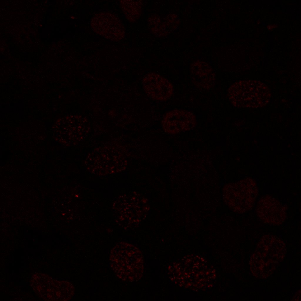
 fixed HepG2 cells using 60097-1-Ig (PCNA antibody), at dilution of 1:200 and CoraLite®488-Conjugated AffiniPure Goat Anti-Mouse IgG(H+L).")
 fixed HepG2 cells using 60097-1-Ig(PCNA antibody) at dilution of 1:100 and Alexa Fluor 488-conjugated AffiniPure Goat Anti-Mouse IgG(H+L).")
 and PCNA (red, Cat. No 60097-1-Ig). Desmin was conjugated to CoraLite-488 fluorescent dye and stains cardiomyocytes (heart muscle cells). PCNA stains proliferating cells, which are numerous in the developing heart. In this image, the four chambers of the heart can be easily visualized. Image credit: @Immunofluorescence on Instagram.")
"PCNA Antibodies" Comparison
View side-by-side comparison of PCNA antibodies from other vendors to find the one that best suits your research needs.
Applications testées
| Résultats positifs en WB | cellules HEK-293, cellules 3T3-L1, cellules 4T1, cellules C6, cellules HeLa, cellules HepG2, cellules Jurkat, cellules K-562, cellules NIH/3T3, cellules RAW 264.7, cellules ROS1728 |
| Résultats positifs en IP | cellules HepG2 |
| Résultats positifs en IHC | tissu de gliome humain, tissu d'amygdalite humain, tissu de cancer du foie humain, tissu de cancer du sein humain, tissu ovarien de souris, tissu placentaire humain il est suggéré de démasquer l'antigène avec un tampon de TE buffer pH 9.0; (*) À défaut, 'le démasquage de l'antigène peut être 'effectué avec un tampon citrate pH 6,0. |
| Résultats positifs en IF/ICC | cellules HepG2, tissu cardiaque de souris |
Dilution recommandée
| Application | Dilution |
|---|---|
| Western Blot (WB) | WB : 1:5000-1:50000 |
| Immunoprécipitation (IP) | IP : 0.5-4.0 ug for 1.0-3.0 mg of total protein lysate |
| Immunohistochimie (IHC) | IHC : 1:500-1:2000 |
| Immunofluorescence (IF)/ICC | IF/ICC : 1:250-1:1000 |
| It is recommended that this reagent should be titrated in each testing system to obtain optimal results. | |
| Sample-dependent, check data in validation data gallery | |
Applications publiées
| KD/KO | See 1 publications below |
| WB | See 116 publications below |
| IHC | See 47 publications below |
| IF | See 35 publications below |
| IP | See 2 publications below |
| CoIP | See 1 publications below |
Informations sur le produit
60097-1-Ig cible PCNA dans les applications de WB, IHC, IF/ICC, IP, CoIP, ELISA et montre une réactivité avec des échantillons Humain, porc, rat, souris
| Réactivité | Humain, porc, rat, souris |
| Réactivité citée | rat, Humain, Lapin, poisson-zèbre, porc, souris |
| Hôte / Isotype | Mouse / IgG1 |
| Clonalité | Monoclonal |
| Type | Anticorps |
| Immunogène | PCNA Protéine recombinante Ag7416 |
| Nom complet | proliferating cell nuclear antigen |
| Masse moléculaire calculée | 29 kDa/31 kDa |
| Poids moléculaire observé | 36-38 kDa |
| Numéro d’acquisition GenBank | BC000491 |
| Symbole du gène | PCNA |
| Identification du gène (NCBI) | 5111 |
| Conjugaison | Non conjugué |
| Forme | Liquide |
| Méthode de purification | Purification par protéine G |
| Tampon de stockage | PBS with 0.02% sodium azide and 50% glycerol |
| Conditions de stockage | Stocker à -20°C. Stable pendant un an après l'expédition. L'aliquotage n'est pas nécessaire pour le stockage à -20oC Les 20ul contiennent 0,1% de BSA. |
Informations générales
Proliferating Cell Nuclear Antigen, commonly known as PCNA, is a protein that acts as a processivity factor for DNA polymerase δ in eukaryotic cells. This protein is an auxiliary protein of DNA polymerase delta and is involved in the control of eukaryotic DNA replication by increasing the polymerase's processibility during elongation of the leading strand. PCNA induces a robust stimulatory effect on the 3'-5' exonuclease and 3'-phosphodiesterase, but not apurinic-apyrimidinic (AP) endonuclease, APEX2 activities. It has to be loaded onto DNA in order to be able to stimulate APEX2. PCNA protein is highly conserved during evolution; the deduced amino acid sequences of rat and human differ by only 4 of 261 amino acids. PCNA has been used as loading control for proliferating cells. The calculated molecular weight of PCNA is 29 kDa, but modified PCNA is 36kDa (PMID: 1358458).
Protocole
| Product Specific Protocols | |
|---|---|
| WB protocol for PCNA antibody 60097-1-Ig | Download protocol |
| IHC protocol for PCNA antibody 60097-1-Ig | Download protocol |
| IF protocol for PCNA antibody 60097-1-Ig | Download protocol |
| IP protocol for PCNA antibody 60097-1-Ig | Download protocol |
| Standard Protocols | |
|---|---|
| Click here to view our Standard Protocols |
Publications
| Species | Application | Title |
|---|---|---|
Nat Commun USP52 acts as a deubiquitinase and promotes histone chaperone ASF1A stabilization. | ||
Nat Commun Cyclophilin J limits inflammation through the blockage of ubiquitin chain sensing. | ||
Microbiome Antioxidant potential of Pediococcus pentosaceus strains from the sow milk bacterial collection in weaned piglets. | ||
Nat Cardiovasc Res Endocardial primary cilia and blood flow regulate EndoMT during endocardial cushion development | ||
Environ Pollut Wnt10a downregulation contributes to MEHP-induced disruption of self-renewal and differentiation balance and proliferation inhibition in GC-1 cells: Insights from multiple transcriptomic profiling | ||
Acta Pharmacol Sin PU.1/Spi1 exacerbates ischemia-reperfusion induced acute kidney injury via upregulating Gata2 and promoting fibroblast activation |
Avis
The reviews below have been submitted by verified Proteintech customers who received an incentive for providing their feedback.
FH Julia (Verified Customer) (08-12-2024) | Really good, specific antibody! Incubated over night at 4C; 1:10000 dilution in 3% milk.
 |
FH Tsimafei (Verified Customer) (08-04-2024) | Excellent antibody. Membrane was incubated at 4C, ON
 |
FH Andrea (Verified Customer) (01-11-2023) | primary antibody were incubated at 4C, ON. Antibody provide a specific signal for mouse Pcna protein
 |
FH Hasan (Verified Customer) (11-04-2022) | U2OS cells fixed with Methanol:Aceton (7:3), 20 min, 4 degrees. Blocked for 1h with 5% FBS in Triton-x 0.1% Stained with the anti-PCNA (60097-1-Ig) (1:200) O/N @4 degress
 |
FH Maria (Verified Customer) (08-07-2021) | PCNA in human primary fibroblasts in culture. Passage number 15-25. 15 ug of protein. 4-15% TGX gel. Blocking BSA 3% in PBST. Primary ab 1:1000 in BSA O/N 4ºC. Secondary ab 1:5000 HRP.
 |

