"PCNA Antibodies" Comparison
View side-by-side comparison of PCNA antibodies from other vendors to find the one that best suits your research needs.
Tested Applications
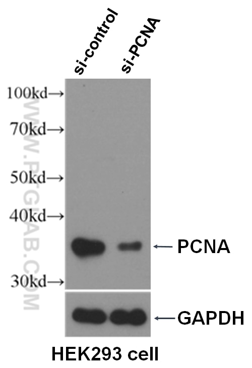
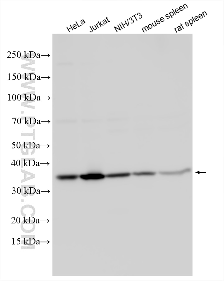
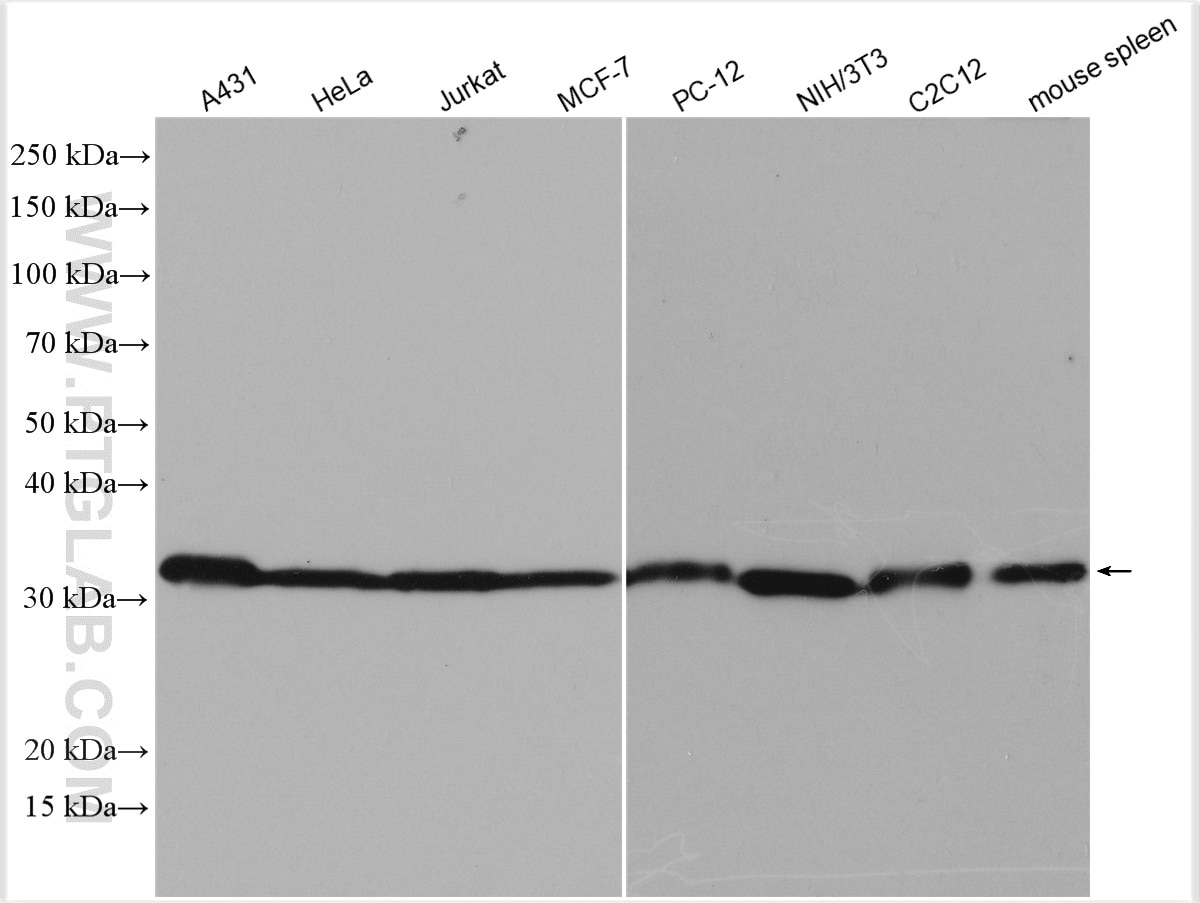
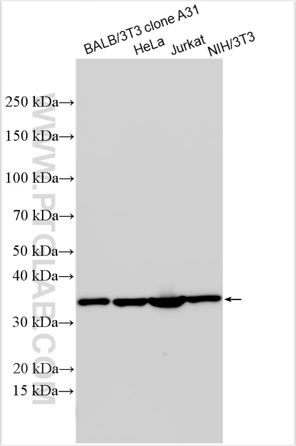
| Positive WB detected in | HeLa cells, A431 cells, BALB/3T3 clone A31 cells, HEK293 cells, Jurkat cells, MCF-7 cells, PC-12 cells, NIH/3T3 cells, C2C12 cells, mouse spleen tissue, rat spleen tissue |
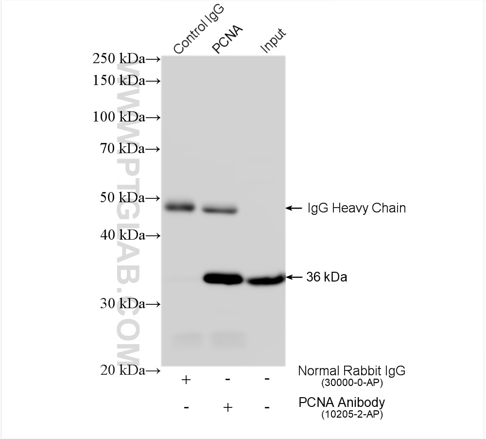
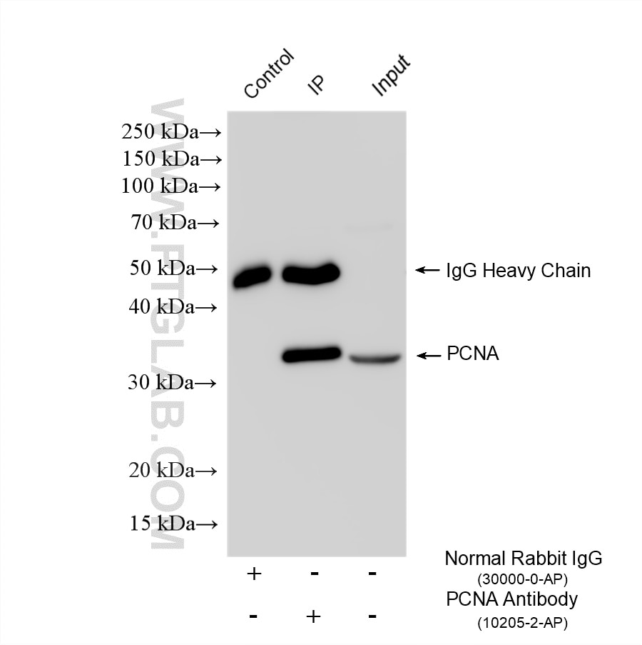
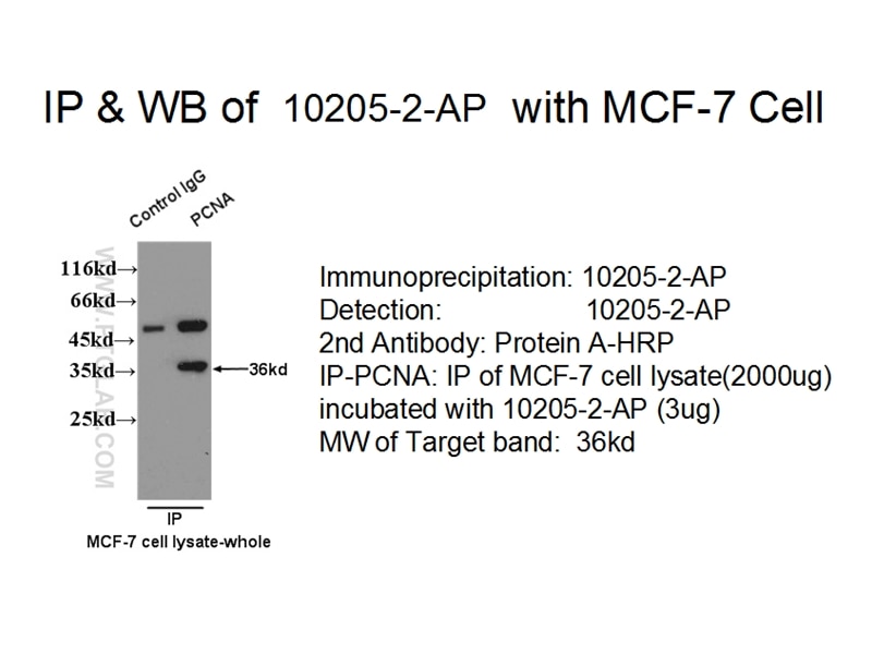
| Positive IP detected in | MCF-7 cells, HeLa cells |
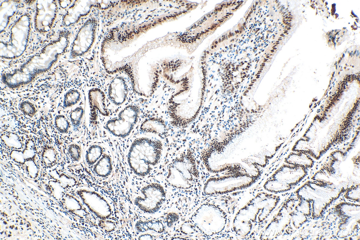
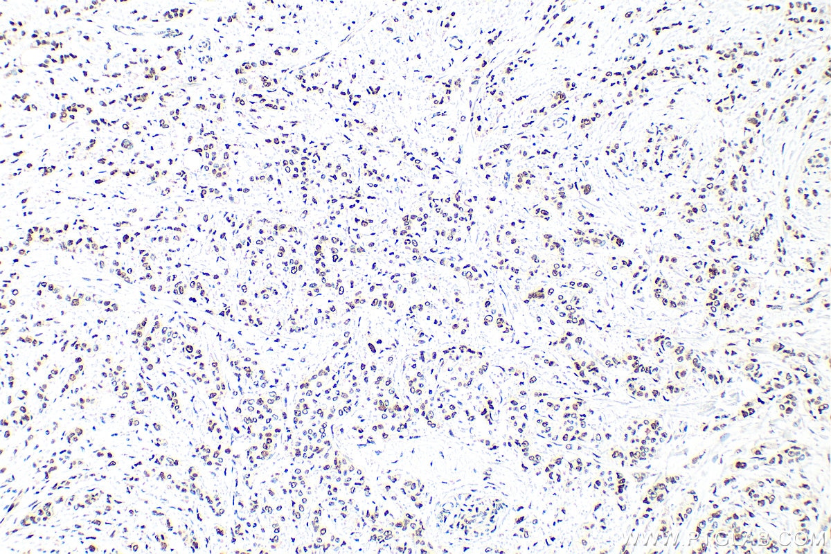
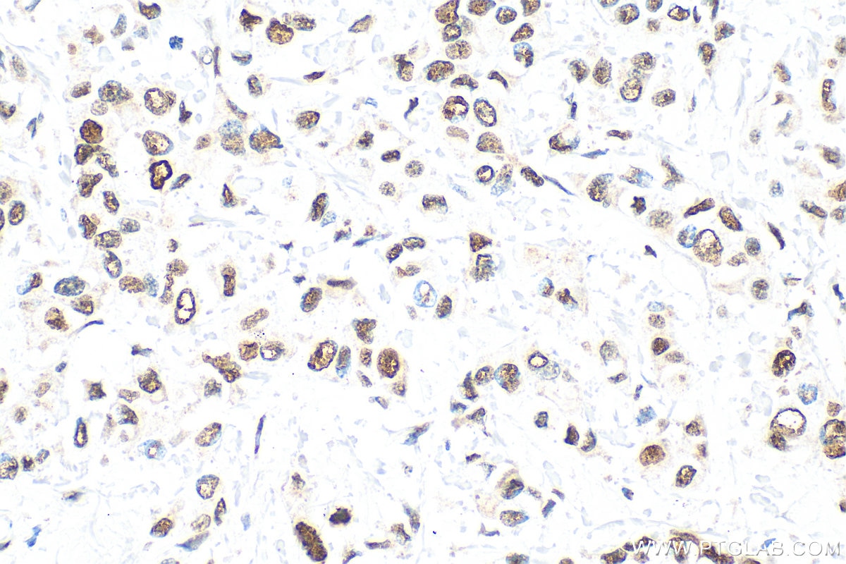
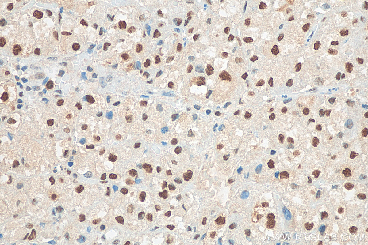
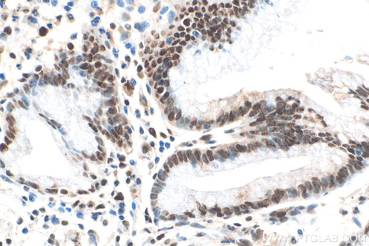
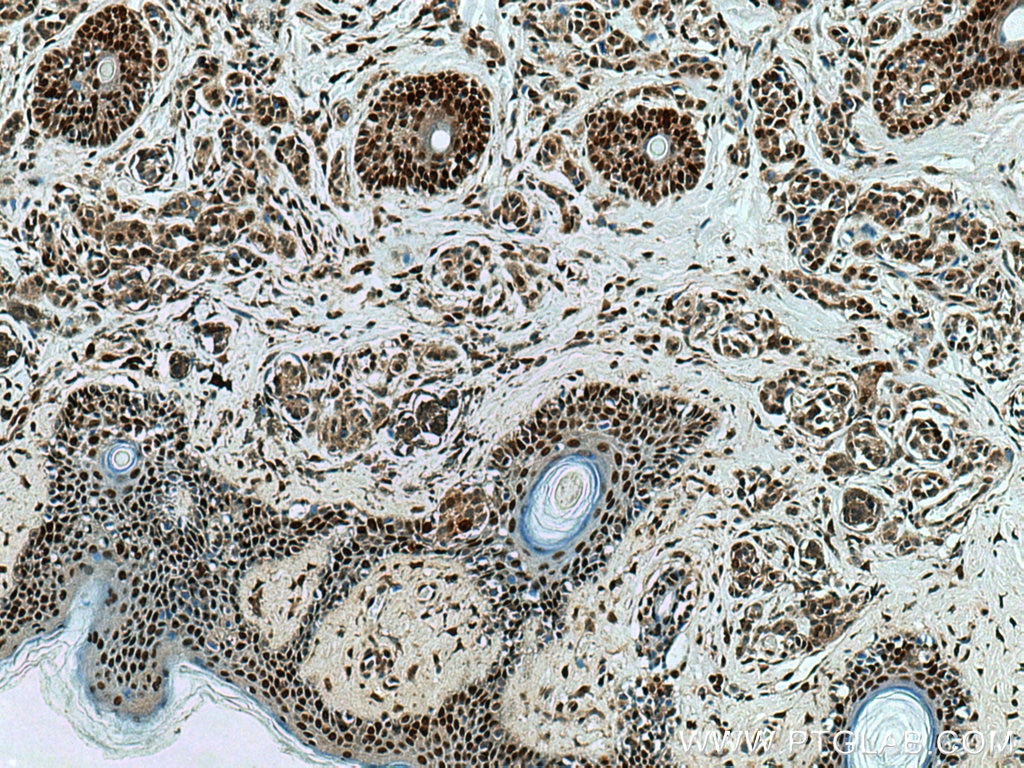
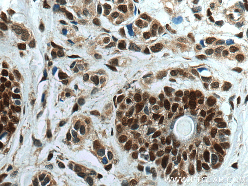
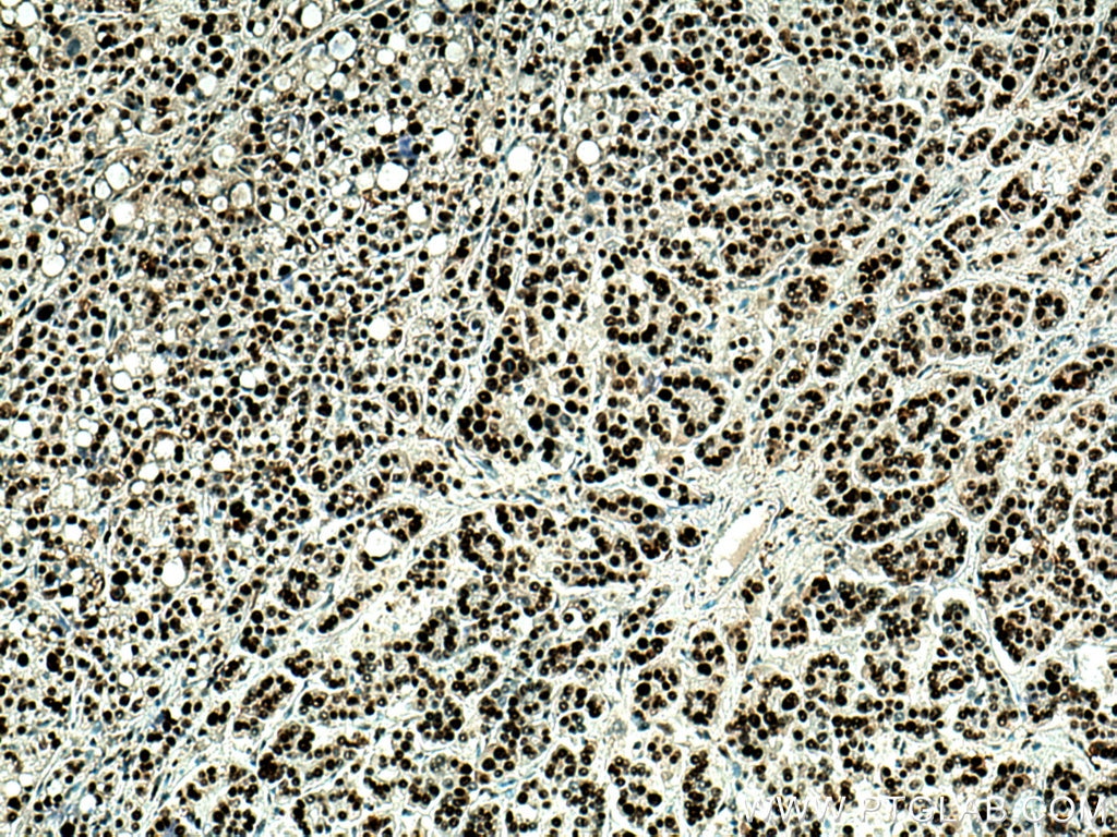
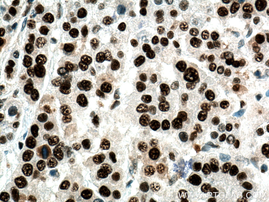
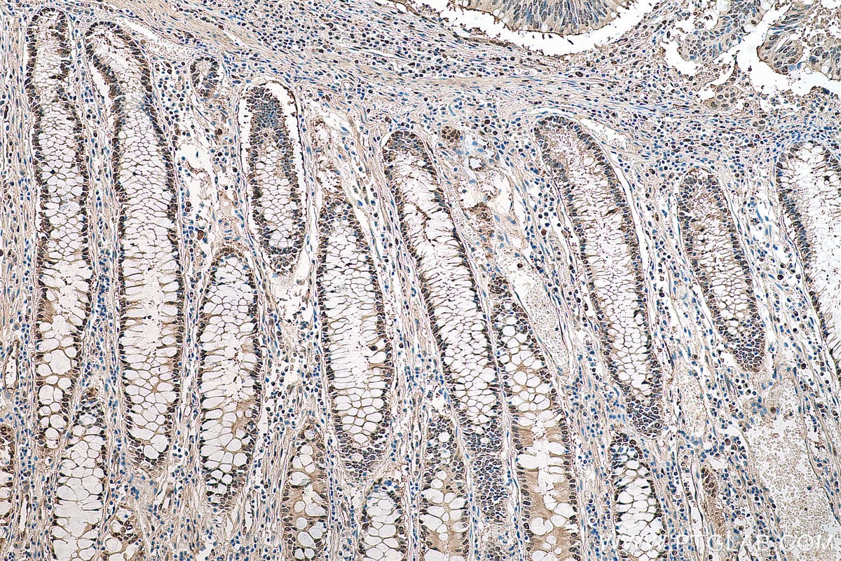
| Positive IHC detected in | human stomach cancer tissue, human breast cancer tissue, human colon cancer tissue, human liver cancer tissue, human malignant melanoma tissue Note: suggested antigen retrieval with TE buffer pH 9.0; (*) Alternatively, antigen retrieval may be performed with citrate buffer pH 6.0 |
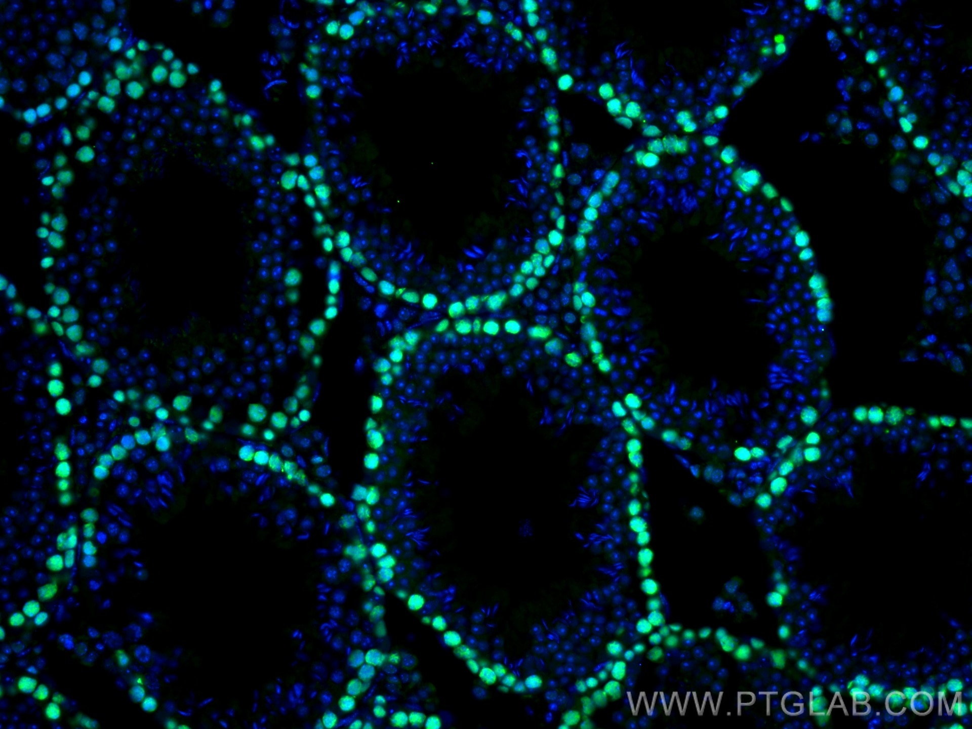
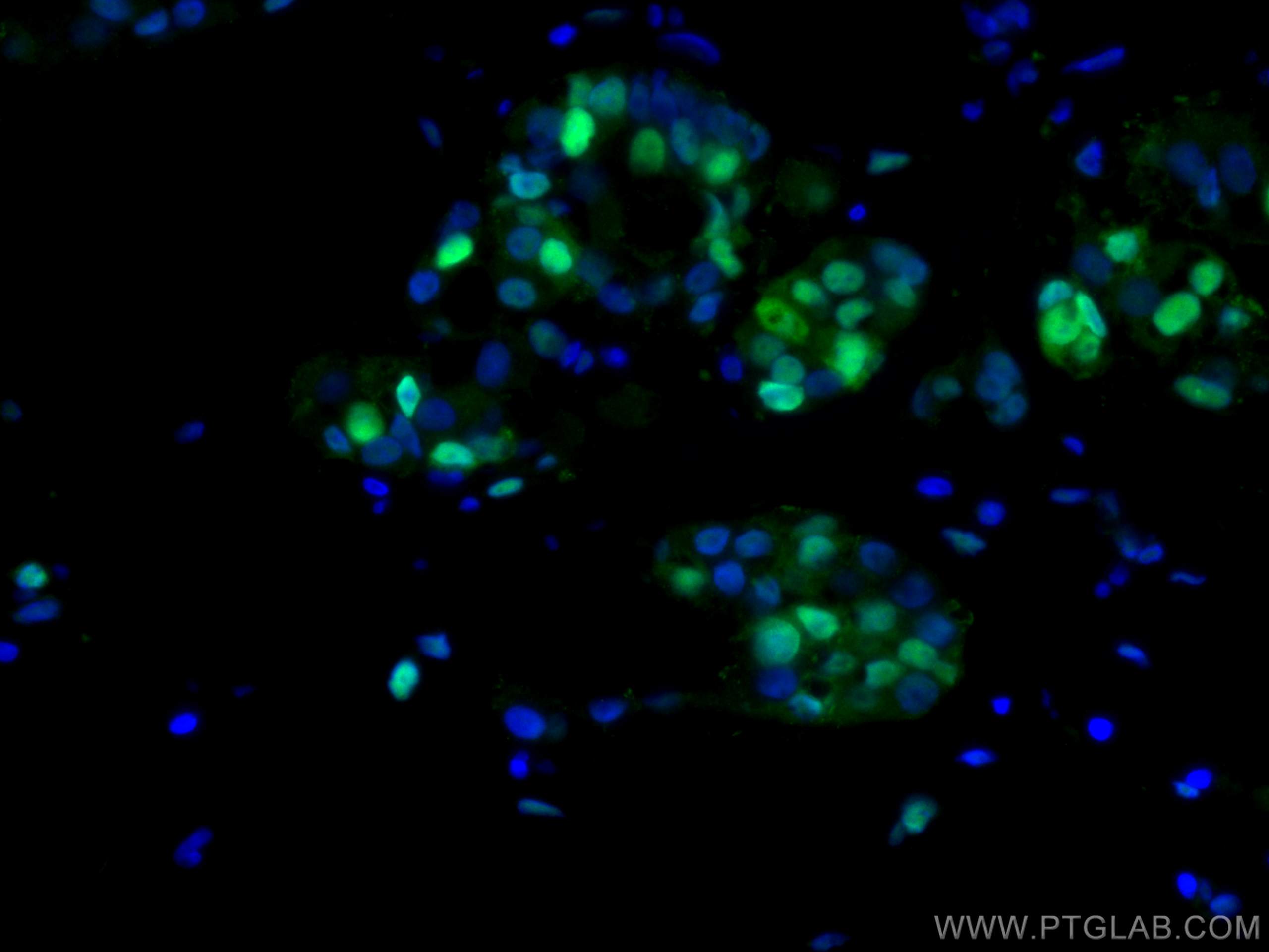
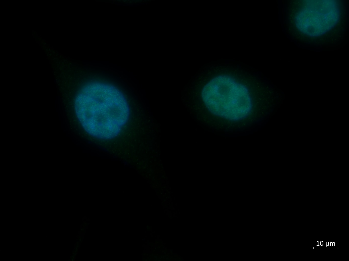
| Positive IF-P detected in | mouse testis tissue, human breast cancer tissue |
Recommended dilution
| Application | Dilution |
|---|---|
| Western Blot (WB) | WB : 1:5000-1:50000 |
| Immunoprecipitation (IP) | IP : 0.5-4.0 ug for 1.0-3.0 mg of total protein lysate |
| Immunohistochemistry (IHC) | IHC : 1:1500-1:6000 |
| Immunofluorescence (IF)-P | IF-P : 1:50-1:500 |
| It is recommended that this reagent should be titrated in each testing system to obtain optimal results. | |
| Sample-dependent, Check data in validation data gallery. | |
Published Applications
| KD/KO | See 6 publications below |
| WB | See 1004 publications below |
| IHC | See 362 publications below |
| IF | See 146 publications below |
| IP | See 5 publications below |
| CoIP | See 2 publications below |
Product Information
10205-2-AP targets PCNA in WB, IHC, IF-P, IP, CoIP, ELISA, Cell treatment applications and shows reactivity with human, mouse, rat samples.
| Tested Reactivity | human, mouse, rat |
| Cited Reactivity | human, mouse, rat, rabbit, chicken, goat, sheep, fish, ducks, medaka embryos |
| Host / Isotype | Rabbit / IgG |
| Class | Polyclonal |
| Type | Antibody |
| Immunogen |
CatNo: Ag0277 Product name: Recombinant human PCNA protein Source: e coli.-derived, PGEX-4T Tag: GST Domain: 8-256 aa of BC000491 Sequence: QGSILKKVLEALKDLINEACWDISSSGVNLQSMDSSHVSLVQLTLRSEGFDTYRCDRNLAMGVNLTSMSKILKCAGNEDIITLRAEDNADTLALVFEAPNQEKVSDYEMKLMDLDVEQLGIPEQEYSCVVKMPSGEFARICRDLSHIGDAVVISCAKDGVKFSASGELGNGNIKLSQTSNVDKEEEAVTIEMNEPVQLTFALRYLNFFTKATPLSSTVTLSMSADVPLVVEYKIADMGHLKYYLAPKIE Predict reactive species |
| Full Name | proliferating cell nuclear antigen |
| Calculated Molecular Weight | 29 kDa/31 kDa |
| Observed Molecular Weight | 36-38 kDa |
| GenBank Accession Number | BC000491 |
| Gene Symbol | PCNA |
| Gene ID (NCBI) | 5111 |
| RRID | AB_2160330 |
| Conjugate | Unconjugated |
| Form | Liquid |
| Purification Method | Antigen affinity purification |
| UNIPROT ID | P12004 |
| Storage Buffer | PBS with 0.02% sodium azide and 50% glycerol, pH 7.3. |
| Storage Conditions | Store at -20°C. Stable for one year after shipment. Aliquoting is unnecessary for -20oC storage. 20ul sizes contain 0.1% BSA. |
Background Information
Proliferating Cell Nuclear Antigen, commonly known as PCNA, is a protein that acts as a processivity factor for DNA polymerase δ in eukaryotic cells. This protein is an auxiliary protein of DNA polymerase delta and is involved in the control of eukaryotic DNA replication by increasing the polymerase's processibility during elongation of the leading strand. PCNA induces a robust stimulatory effect on the 3'-5' exonuclease and 3'-phosphodiesterase, but not apurinic-apyrimidinic (AP) endonuclease, APEX2 activities. It has to be loaded onto DNA in order to be able to stimulate APEX2. PCNA protein is highly conserved during evolution; the deduced amino acid sequences of rat and human differ by only 4 of 261 amino acids. PCNA has been used as loading control for proliferating cells. This antibody is a rabbit polyclonal antibody raised against an internal region of human PCNA. The calculated molecular weight of PCNA is 29 kDa, but modified PCNA is 36kDa (PMID: 1358458).
Protocols
| Product Specific Protocols | |
|---|---|
| IF protocol for PCNA antibody 10205-2-AP | Download protocol |
| IHC protocol for PCNA antibody 10205-2-AP | Download protocol |
| IP protocol for PCNA antibody 10205-2-AP | Download protocol |
| WB protocol for PCNA antibody 10205-2-AP | Download protocol |
| Standard Protocols | |
|---|---|
| Click here to view our Standard Protocols |
Publications
| Species | Application | Title |
|---|---|---|
J Hematol Oncol METTL16 promotes liver cancer stem cell self-renewal via controlling ribosome biogenesis and mRNA translation | ||
Mil Med Res Aspartoacylase suppresses prostate cancer progression by blocking LYN activation | ||
Protein Cell NDFIP1 limits cellular TAZ accumulation via exosomal sorting to inhibit NSCLC proliferation | ||
Brain Behav Immun An enriched environment restores hepatitis B vaccination-mediated impairments in synaptic function through IFN-γ/Arginase1 signaling. | ||
Nat Commun Four-dimensional hydrogel dressing adaptable to the urethral microenvironment for scarless urethral reconstruction | ||
Mol Cell Alu repeat-containing RNAs spatially organize actively transcribed genomic regions around nuclear speckles. |
Reviews
The reviews below have been submitted by verified Proteintech customers who received an incentive for providing their feedback.
FH MALLIKARJUNA (Verified Customer) (10-14-2025) | GOOD FOR IF
|
FH Mi (Verified Customer) (06-25-2023) | It works well in human brown adipocyte cells.
|
FH Emma (Verified Customer) (11-29-2021) | Nice antibody which worked well on PC3 cells fixed with 4% PFA. Antibody used at 1:50
 |