- Phare
- Validé par KD/KO
Anticorps Polyclonal de lapin anti-PHLDA1
PHLDA1 Polyclonal Antibody for WB, IHC, IF/ICC, ELISA
Hôte / Isotype
Lapin / IgG
Réactivité testée
Humain, souris
Applications
WB, IHC, IF/ICC, ELISA
Conjugaison
Non conjugué
N° de cat : 18263-1-AP
Synonymes
Galerie de données de validation
 at dilution of 1:4000 incubated at room temperature for 1.5 hours.")
 at dilution of 1:600 incubated at room temperature for 1.5 hours.")
 at dilution of 1:800 (under 20x lens). Heat mediated antigen retrieval with Tris-EDTA buffer (pH 9.0).")
 fixed A375 cells using PHLDA1 antibody (18263-1-AP) at dilution of 1:400 and CoraLite®488-Conjugated AffiniPure Goat Anti-Rabbit IgG(H+L).")
Applications testées
| Résultats positifs en WB | cellules A375, cellules BxPC-3, cellules HeLa, cellules MDA-MB-231, tissu cérébral de souris |
| Résultats positifs en IHC | tissu de cancer de la peau humain, il est suggéré de démasquer l'antigène avec un tampon de TE buffer pH 9.0; (*) À défaut, 'le démasquage de l'antigène peut être 'effectué avec un tampon citrate pH 6,0. |
| Résultats positifs en IF/ICC | cellules A375, |
Dilution recommandée
| Application | Dilution |
|---|---|
| Western Blot (WB) | WB : 1:1000-1:6000 |
| Immunohistochimie (IHC) | IHC : 1:400-1:1600 |
| Immunofluorescence (IF)/ICC | IF/ICC : 1:200-1:800 |
| It is recommended that this reagent should be titrated in each testing system to obtain optimal results. | |
| Sample-dependent, check data in validation data gallery | |
Applications publiées
| KD/KO | See 6 publications below |
| WB | See 9 publications below |
| IHC | See 2 publications below |
| IF | See 2 publications below |
Informations sur le produit
18263-1-AP cible PHLDA1 dans les applications de WB, IHC, IF/ICC, ELISA et montre une réactivité avec des échantillons Humain, souris
| Réactivité | Humain, souris |
| Réactivité citée | Humain, souris |
| Hôte / Isotype | Lapin / IgG |
| Clonalité | Polyclonal |
| Type | Anticorps |
| Immunogène | PHLDA1 Protéine recombinante Ag13125 |
| Nom complet | pleckstrin homology-like domain, family A, member 1 |
| Masse moléculaire calculée | 45 kDa |
| Poids moléculaire observé | 40-45 kDa |
| Numéro d’acquisition GenBank | BC018929 |
| Symbole du gène | PHLDA1 |
| Identification du gène (NCBI) | 22822 |
| Conjugaison | Non conjugué |
| Forme | Liquide |
| Méthode de purification | Purification par affinité contre l'antigène |
| Tampon de stockage | PBS with 0.02% sodium azide and 50% glycerol |
| Conditions de stockage | Stocker à -20°C. Stable pendant un an après l'expédition. L'aliquotage n'est pas nécessaire pour le stockage à -20oC Les 20ul contiennent 0,1% de BSA. |
Informations générales
PHLDA1, also known as PHRIP and TDAG51, is a multifunctional protein involved in various biological processes. It can induce apoptosis in various cell types, including T cells, hippocampal cells, endothelial cells, melanoma cells, and mouse embryonic fibroblasts(PMID: 30207029). PHLDA1 plays a role in inhibiting growth factor signaling and has been implicated in tumor suppression through its ability to repress Akt activity by binding to phosphatidylinositol (PIP) lipids(PMID: 36142223).
Protocole
| Product Specific Protocols | |
|---|---|
| WB protocol for PHLDA1 antibody 18263-1-AP | Download protocol |
| IHC protocol for PHLDA1 antibody 18263-1-AP | Download protocol |
| IF protocol for PHLDA1 antibody 18263-1-AP | Download protocol |
| Standard Protocols | |
|---|---|
| Click here to view our Standard Protocols |
Publications
| Species | Application | Title |
|---|---|---|
Brain Behav Immun PHLDA1 promotes microglia-mediated neuroinflammation via regulating K63-linked ubiquitination of TRAF6.
| ||
Inflammation Multiple Machine Learning Identifies Key Gene PHLDA1 Suppressing NAFLD Progression | ||
Life Sci PHLDA1 is a new therapeutic target of oxidative stress and ischemia reperfusion-induced myocardial injury.
| ||
Inflammation TDAG51-Deficiency Podocytes are Protected from High-Glucose-Induced Damage Through Nrf2 Activation via the AKT-GSK-3β Pathway.
| ||
Hum Exp Toxicol Loss of PHLDA1 has a protective role in OGD/R-injured neurons via regulation of the GSK-3β/Nrf2 pathway.
| ||
Neuroreport Egr1 promotes Nlrc4-dependent neuronal pyroptosis through phlda1 in an in-vitro model of intracerebral hemorrhage
|
Avis
The reviews below have been submitted by verified Proteintech customers who received an incentive for providing their feedback.
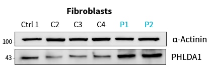
FH Lukas (Verified Customer) (01-31-2025) | worked very well for Western blot in patient primary fibroblasts and cell lines (in 1% milk at +4°C overnight)
 |

