- Phare
- Validé par KD/KO
Anticorps Polyclonal de lapin anti-PIBF1
PIBF1 Polyclonal Antibody for WB, IHC, IF/ICC, IP, ELISA
Hôte / Isotype
Lapin / IgG
Réactivité testée
Humain
Applications
WB, IHC, IF/ICC, IP, ELISA
Conjugaison
Non conjugué
N° de cat : 14413-1-AP
Synonymes
Galerie de données de validation
 at dilution of 1:3000 incubated at room temperature for 1.5 hours.")
 at dilution of 1:1000 incubated at room temperature for 1.5 hours.")
 at dilution of 1:500 incubated at room temperature for 1.5 hours.")
 at dilution of 1:500 incubated at room temperature for 1.5 hours.")
 with HEK-293 cells lysate 1400 ug.")
 at dilution of 1:200 (under 20x lens). Heat mediated antigen retrieval with Tris-EDTA buffer (pH 9.0).")
 fixed HEK-293 cells using PIBF1 antibody (14413-1-AP) at dilution of 1:400 and CoraLite®488-Conjugated Goat Anti-Rabbit IgG(H+L), Gamma Tubulin antibody (66320-1-Ig, Clone: 3F9H8, red).")
Applications testées
| Résultats positifs en WB | cellules HEK-293, cellules HeLa, cellules K-562, cellules MCF-7 |
| Résultats positifs en IP | cellules HEK-293, |
| Résultats positifs en IHC | human intrahepatic cholangiocarcinoma tissue, il est suggéré de démasquer l'antigène avec un tampon de TE buffer pH 9.0; (*) À défaut, 'le démasquage de l'antigène peut être 'effectué avec un tampon citrate pH 6,0. |
| Résultats positifs en IF/ICC | cellules HEK-293, |
Dilution recommandée
| Application | Dilution |
|---|---|
| Western Blot (WB) | WB : 1:1000-1:6000 |
| Immunoprécipitation (IP) | IP : 0.5-4.0 ug for 1.0-3.0 mg of total protein lysate |
| Immunohistochimie (IHC) | IHC : 1:50-1:500 |
| Immunofluorescence (IF)/ICC | IF/ICC : 1:200-1:800 |
| It is recommended that this reagent should be titrated in each testing system to obtain optimal results. | |
| Sample-dependent, check data in validation data gallery | |
Applications publiées
| KD/KO | See 3 publications below |
| WB | See 2 publications below |
| IF | See 6 publications below |
| IP | See 1 publications below |
Informations sur le produit
14413-1-AP cible PIBF1 dans les applications de WB, IHC, IF/ICC, IP, ELISA et montre une réactivité avec des échantillons Humain
| Réactivité | Humain |
| Réactivité citée | Humain |
| Hôte / Isotype | Lapin / IgG |
| Clonalité | Polyclonal |
| Type | Anticorps |
| Immunogène | PIBF1 Protéine recombinante Ag5755 |
| Nom complet | progesterone immunomodulatory binding factor 1 |
| Masse moléculaire calculée | 90 kDa |
| Poids moléculaire observé | 90 kDa |
| Numéro d’acquisition GenBank | BC051911 |
| Symbole du gène | PIBF1 |
| Identification du gène (NCBI) | 10464 |
| Conjugaison | Non conjugué |
| Forme | Liquide |
| Méthode de purification | Purification par affinité contre l'antigène |
| Tampon de stockage | PBS with 0.02% sodium azide and 50% glycerol |
| Conditions de stockage | Stocker à -20°C. Stable pendant un an après l'expédition. L'aliquotage n'est pas nécessaire pour le stockage à -20oC Les 20ul contiennent 0,1% de BSA. |
Informations générales
PIBF1 is induced by the steroid hormone progesterone and plays a role in the maintenance of pregnancy. PIBF1 regulates multiple facets of the immune system to promote normal pregnancy including cytokine synthesis, natural killer (NK) cell activity, and arachidonic acid metabolism. Low serum levels of this protein have been associated with spontaneous pre-term labor in humans. PIBF1 may promote the proliferation, migration and invasion of glioma.
Protocole
| Product Specific Protocols | |
|---|---|
| WB protocol for PIBF1 antibody 14413-1-AP | Download protocol |
| IHC protocol for PIBF1 antibody 14413-1-AP | Download protocol |
| IF protocol for PIBF1 antibody 14413-1-AP | Download protocol |
| IP protocol for PIBF1 antibody 14413-1-AP | Download protocol |
| Standard Protocols | |
|---|---|
| Click here to view our Standard Protocols |
Publications
| Species | Application | Title |
|---|---|---|
Elife Centriolar satellites assemble centrosomal microcephaly proteins to recruit CDK2 and promote centriole duplication.
| ||
J Cell Biol A ciliopathy complex builds distal appendages to initiate ciliogenesis.
| ||
EMBO Rep Zika virus alters centrosome organization to suppress the innate immune response. | ||
PLoS Biol The evolutionary conserved proteins CEP90, FOPNL, and OFD1 recruit centriolar distal appendage proteins to initiate their assembly
| ||
Cell Rep CCDC57 Cooperates with Microtubules and Microcephaly Protein CEP63 and Regulates Centriole Duplication and Mitotic Progression. | ||
EMBO J Human SFI1 and Centrin form a complex critical for centriole architecture and ciliogenesis |
Avis
The reviews below have been submitted by verified Proteintech customers who received an incentive for providing their feedback.
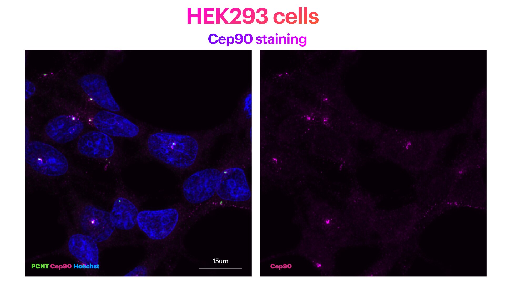
FH Elisa (Verified Customer) (06-20-2022) | HEK293 cells stained for Hoechst (DNA marker, in green), Cep90 (mother centriole distal appendage marker, in magenta) and PCNT (pericentriolar matrix marker, in green). HEK293 cells were plated on Poly-lysine coated coverslips and fixed in cold methanol for 2' at -20C. Cells were then rehydrated with PBS for 5'. Membrane permeabilisation was then performed with 0.1% Triton + 0.1% Tween +0.01%SDS in PBS for 5'. Cells were finally incubated with blocking buffer (5% BSA+ 0.1% Tween in PBS) for 30' at RT. Primary antibody was diluted in blocking buffer 1:200 and incubated for 1h at room temperature. Alexa-555-Anti-rabbit was used as secondary antibody (1:600 dilution) (1h at room temperature). Cep90 antibody recognises clearly dots at the centrosome (identified by the presence of PCNT).
 |
FH Pierrick (Verified Customer) (10-24-2019) | Antibody mostly used in wester blot and work well at 1/1000 dilution on RPE1 and HEK293 sample
|

