- Phare
- Validé par KD/KO
Anticorps Polyclonal de lapin anti-PML
PML Polyclonal Antibody for WB, IHC, IF/ICC, IP, ELISA
Hôte / Isotype
Lapin / IgG
Réactivité testée
Humain
Applications
WB, IHC, IF/ICC, IP, ELISA
Conjugaison
Non conjugué
N° de cat : 21041-1-AP
Synonymes
Galerie de données de validation
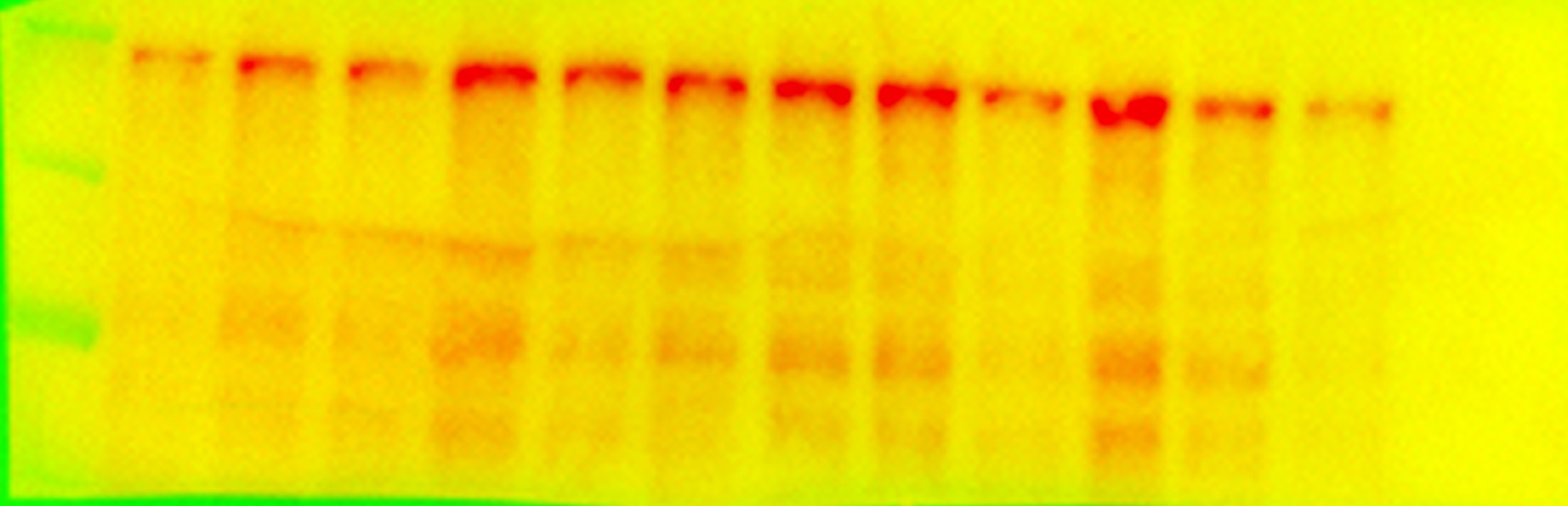
 with sh-Control and sh-PML transfected HeLa cells.")
 at dilution of 1:300 incubated at room temperature for 1.5 hours.")
 with HEK-293 cells lysate 3200ug.")
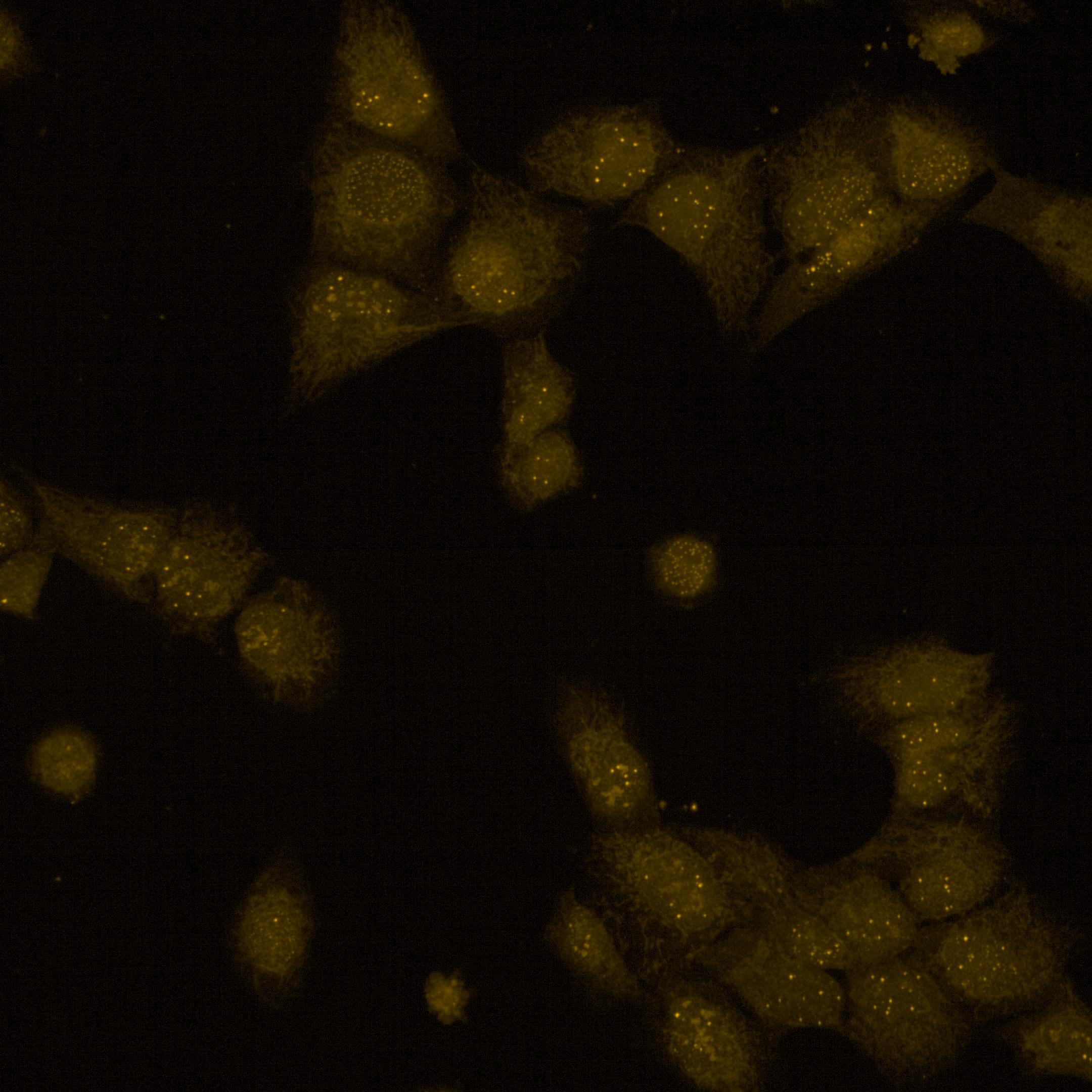
 at dilution of 1:200 (under 10x lens).")
 at dilution of 1:200 (under 40x lens).")
 fixed HeLa cells using 21041-1-AP (PML antibody) at dilution of 1:50 and Alexa Fluor 488-conjugated AffiniPure Goat Anti-Rabbit IgG(H+L).")
 fixed HeLa cells using PML antibody (21041-1-AP) at dilution of 1:200 and CoraLite®488-Conjugated AffiniPure Goat Anti-Rabbit IgG(H+L), CL594-phalloidin (red).")
 fixed HeLa cells using PML antibody (21041-1-AP) at dilution of 1:200 and CoraLite®488-Conjugated AffiniPure Goat Anti-Rabbit IgG(H+L), CL594-Phalloidin (red).")
Applications testées
| Résultats positifs en WB | cellules HeLa, cellules HEK-293 |
| Résultats positifs en IP | cellules HEK-293 |
| Résultats positifs en IHC | tissu hépatique humain il est suggéré de démasquer l'antigène avec un tampon de TE buffer pH 9.0; (*) À défaut, 'le démasquage de l'antigène peut être 'effectué avec un tampon citrate pH 6,0. |
| Résultats positifs en IF/ICC | cellules HeLa, |
Dilution recommandée
| Application | Dilution |
|---|---|
| Western Blot (WB) | WB : 1:1000-1:4000 |
| Immunoprécipitation (IP) | IP : 0.5-4.0 ug for 1.0-3.0 mg of total protein lysate |
| Immunohistochimie (IHC) | IHC : 1:100-1:400 |
| Immunofluorescence (IF)/ICC | IF/ICC : 1:50-1:500 |
| It is recommended that this reagent should be titrated in each testing system to obtain optimal results. | |
| Sample-dependent, check data in validation data gallery | |
Applications publiées
| KD/KO | See 2 publications below |
| WB | See 13 publications below |
| IHC | See 1 publications below |
| IF | See 5 publications below |
Informations sur le produit
21041-1-AP cible PML dans les applications de WB, IHC, IF/ICC, IP, ELISA et montre une réactivité avec des échantillons Humain
| Réactivité | Humain |
| Réactivité citée | Humain |
| Hôte / Isotype | Lapin / IgG |
| Clonalité | Polyclonal |
| Type | Anticorps |
| Immunogène | PML Protéine recombinante Ag15377 |
| Nom complet | promyelocytic leukemia |
| Masse moléculaire calculée | 882 aa, 98 kDa |
| Poids moléculaire observé | 120 kDa |
| Numéro d’acquisition GenBank | BC020994 |
| Symbole du gène | PML |
| Identification du gène (NCBI) | 5371 |
| Conjugaison | Non conjugué |
| Forme | Liquide |
| Méthode de purification | Purification par affinité contre l'antigène |
| Tampon de stockage | PBS with 0.02% sodium azide and 50% glycerol |
| Conditions de stockage | Stocker à -20°C. Stable pendant un an après l'expédition. L'aliquotage n'est pas nécessaire pour le stockage à -20oC Les 20ul contiennent 0,1% de BSA. |
Informations générales
PML, also named as MYL, RNF71 and TRIM19, is a member of RBCC/TRIM family of proteins that possess a RING finger domain, B-box, and coiled-coil domain. It functions as tumor suppressor. PML is required for normal, caspase-dependent apoptosis in response to DNA damage, FAS, TNF, or interferons. It plays a role in transcription regulation, DNA damage response, DNA repair and chromatin organization. PML plays a role in processes regulated by retinoic acid, regulation of cell division, terminal differentiation of myeloid precursor cells and differentiation of neural progenitor cells. It regulates PTEN compartmentalization through the inhibition of USP7-mediated deubiquitinylation. A large number of alternative spliced transcripts are synthesized from the PML gene, resulting in a variety of PML proteins ranging in molecular weight from 48-97 kDa (PMID:11704850). And it can be detected as 70-130 kDa or larger due to the modification (especially SUMO) (PMID: 16778193, 22438555).
Protocole
| Product Specific Protocols | |
|---|---|
| WB protocol for PML antibody 21041-1-AP | Download protocol |
| IHC protocol for PML antibody 21041-1-AP | Download protocol |
| IF protocol for PML antibody 21041-1-AP | Download protocol |
| IP protocol for PML antibody 21041-1-AP | Download protocol |
| Standard Protocols | |
|---|---|
| Click here to view our Standard Protocols |
Publications
| Species | Application | Title |
|---|---|---|
Nucleic Acids Res mRNAs are sorted for export or degradation before passing through nuclear speckles. | ||
Theranostics Autophagic degradation of PML promotes susceptibility to HSV-1 by stress-induced corticosterone. | ||
Front Oncol Identification of a Two-Gene (PML-EPB41) Signature With Independent Prognostic Value in Osteosarcoma. | ||
J Ginseng Res 20(S)-ginsenoside Rh2 induces caspase-dependent promyelocytic leukemia-retinoic acid receptor A degradation in NB4 cells via Akt/Bax/caspase9 and TNF-α/caspase8 signaling cascades. |
Avis
The reviews below have been submitted by verified Proteintech customers who received an incentive for providing their feedback.
FH Ipsita (Verified Customer) (07-23-2025) | Antibody works great for mouse tissue samples as well as adherent cells like Hela and Human Corneal Epithelial cells
|
FH Tatyana (Verified Customer) (03-11-2023) | The antibody works really well in ICC in human cells (in 5% goat serum in PBST, ON incubation).
 |
FH Eishi (Verified Customer) (11-17-2017) | Dear colleagues,find attached the WB results. Experimental conditions:Whole protein extraction from mouse embryonic stem cells 25ug protein/laneTransfer 1h30min, 100V, 4C1AB incubation: o/n 4C, 1:500 dilution in milk2AB incubation: 1h RT, 1:20000 dilution in milkDevelopment with femtoThe observed MW is much higher than the expected, with a strong band around 200KDa. This raises the question if the antibody binds specifically to our protein of interest
 |

