- Phare
- Validé par KD/KO
Anticorps Polyclonal de lapin anti-Pinin
Pinin Polyclonal Antibody for WB, IF/ICC, IP, ELISA
Hôte / Isotype
Lapin / IgG
Réactivité testée
Humain et plus (1)
Applications
WB, IHC, IF/ICC, IP, RIP, ELISA
Conjugaison
Non conjugué
N° de cat : 18266-1-AP
Synonymes
Galerie de données de validation
 at dilution of 1:1000 incubated at room temperature for 1.5 hours.")
 with HeLa cells lysate 520ug.")
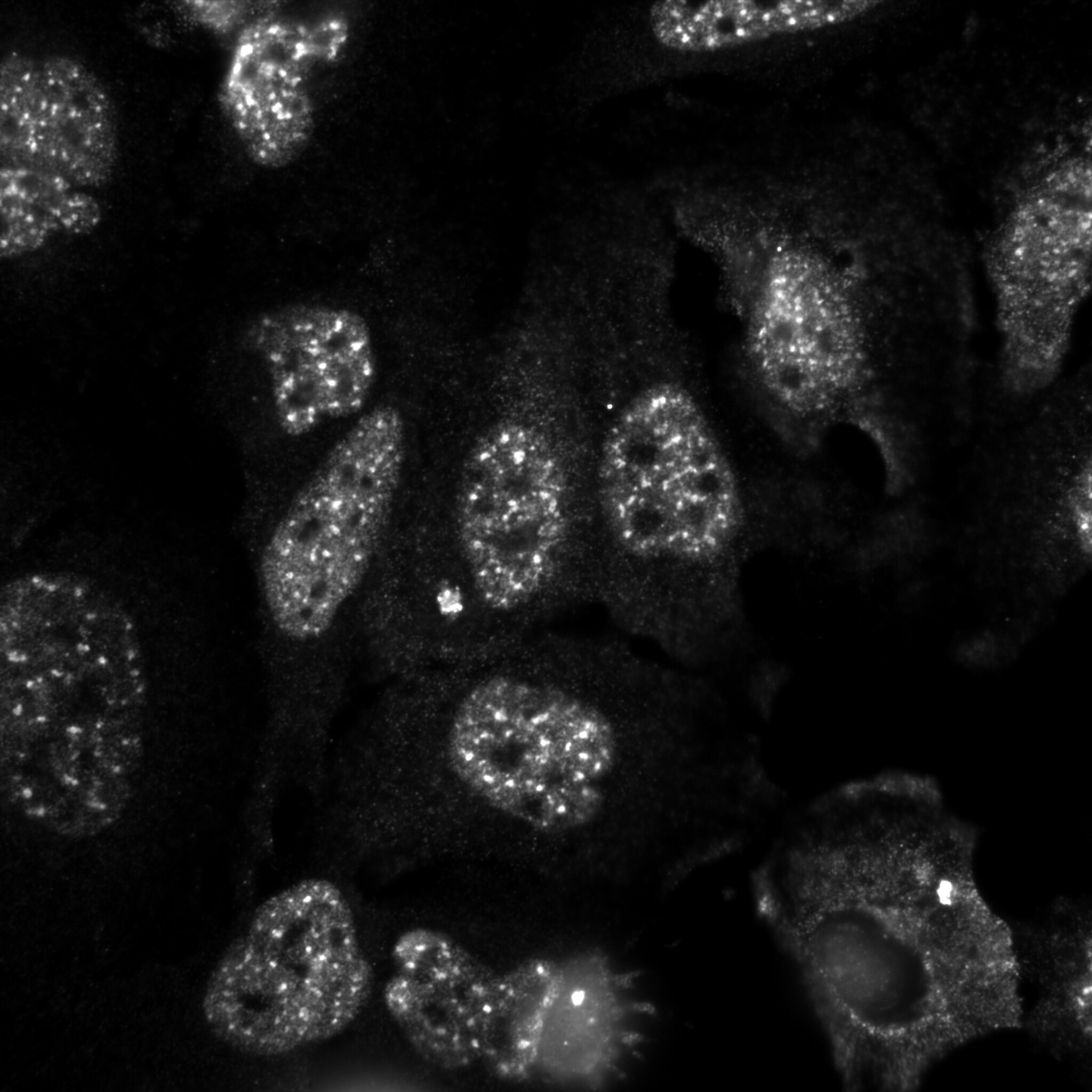
 fixed HeLa cells using Pinin antibody (18266-1-AP) at dilution of 1:200 and CoraLite®488-Conjugated AffiniPure Goat Anti-Rabbit IgG(H+L).")
 fixed HeLa cells using Pinin antibody (18266-1-AP) at dilution of 1:600 and Multi-rAb CoraLite ® Plus 488-Goat Anti-Rabbit Recombinant Secondary Antibody (H+L) (RGAR002), CL594-phalloidin (red).")
Applications testées
| Résultats positifs en WB | cellules HeLa, cellules HepG2 |
| Résultats positifs en IP | cellules HeLa |
| Résultats positifs en IF/ICC | cellules HeLa, |
Dilution recommandée
| Application | Dilution |
|---|---|
| Western Blot (WB) | WB : 1:500-1:2000 |
| Immunoprécipitation (IP) | IP : 0.5-4.0 ug for 1.0-3.0 mg of total protein lysate |
| Immunofluorescence (IF)/ICC | IF/ICC : 1:300-1:1200 |
| It is recommended that this reagent should be titrated in each testing system to obtain optimal results. | |
| Sample-dependent, check data in validation data gallery | |
Applications publiées
| KD/KO | See 1 publications below |
| WB | See 5 publications below |
| IHC | See 1 publications below |
| IF | See 3 publications below |
| IP | See 1 publications below |
| RIP | See 1 publications below |
Informations sur le produit
18266-1-AP cible Pinin dans les applications de WB, IHC, IF/ICC, IP, RIP, ELISA et montre une réactivité avec des échantillons Humain
| Réactivité | Humain |
| Réactivité citée | Humain, souris |
| Hôte / Isotype | Lapin / IgG |
| Clonalité | Polyclonal |
| Type | Anticorps |
| Immunogène | Pinin Protéine recombinante Ag6643 |
| Nom complet | pinin, desmosome associated protein |
| Masse moléculaire calculée | 82 kDa |
| Poids moléculaire observé | 82 kDa, 140 kDa |
| Numéro d’acquisition GenBank | BC062602 |
| Symbole du gène | Pinin |
| Identification du gène (NCBI) | 5411 |
| Conjugaison | Non conjugué |
| Forme | Liquide |
| Méthode de purification | Purification par affinité contre l'antigène |
| Tampon de stockage | PBS with 0.02% sodium azide and 50% glycerol |
| Conditions de stockage | Stocker à -20°C. Stable pendant un an après l'expédition. L'aliquotage n'est pas nécessaire pour le stockage à -20oC Les 20ul contiennent 0,1% de BSA. |
Informations générales
PNN, also named as DRS or 140 kDa nuclear and cell adhesion-related phosphoprotein, is a 717 amino acid protein, which belongs to the pinin family. PNN localizes in the plasma membrane and is expressed in placenta, lung, liver, kidney, pancreas, spleen, thymus, prostate, testis, ovary, small intestine, colon, heart, epidermis, esophagus, brain and smooth and skeletal muscle. PNN is expressed strongly in melanoma metastasis lesions and advanced primary tumors. PNN as a transcriptional activator binds to the E-box 1 core sequence of the E-cadherin promoter gene. PNN is involved in the establishment and maintenance of epithelia cell-cell adhesion. PNN is a potential tumor suppressor for renal cell carcinoma. The calculated molecular weight of PNN is 82 kDa (native form), but the post-modified protein is 140 kDa (phospho form of isoform 1).
Protocole
| Product Specific Protocols | |
|---|---|
| WB protocol for Pinin antibody 18266-1-AP | Download protocol |
| IF protocol for Pinin antibody 18266-1-AP | Download protocol |
| IP protocol for Pinin antibody 18266-1-AP | Download protocol |
| Standard Protocols | |
|---|---|
| Click here to view our Standard Protocols |
Publications
| Species | Application | Title |
|---|---|---|
Cell Metab NEAT1 is essential for metabolic changes that promote breast cancer growth and metastasis. | ||
Mol Oncol LncRNA AATBC regulates Pinin to promote metastasis in nasopharyngeal carcinoma.
| ||
Oncotarget Quantitative proteomics reveals molecular mechanism of gamabufotalin and its potential inhibition on Hsp90 in lung cancer. | ||
Exp Physiol Methyltransferase like 3 enhances pinin mRNA stability through N6 -methyladenosine modification to augment tumourigenesis of colon adenocarcinoma | ||
Oncotarget Pinin associates with prognosis of hepatocellular carcinoma through promoting cell proliferation and suppressing glucose deprivation-induced apoptosis. | ||
Cell Rep Stress-induced TDP-43 nuclear condensation causes splicing loss of function and STMN2 depletion |
Avis
The reviews below have been submitted by verified Proteintech customers who received an incentive for providing their feedback.
FH Tatyana (Verified Customer) (05-14-2023) | ICC on HeLa cells; antibody diluted in 5% goat serum in PBST; 2 h at RT temperature incubation. Labels nuclear speckles. Very good signal
 |

