- Phare
- Validé par KD/KO
Anticorps Polyclonal de lapin anti-PROX1
PROX1 Polyclonal Antibody for WB, IF/ICC, FC (Intra), IP, ELISA
Hôte / Isotype
Lapin / IgG
Réactivité testée
Humain et plus (4)
Applications
WB, IF/ICC, FC (Intra), IP, ChIP, ELISA
Conjugaison
Non conjugué
N° de cat : 11067-2-AP
Synonymes
Galerie de données de validation
Applications testées
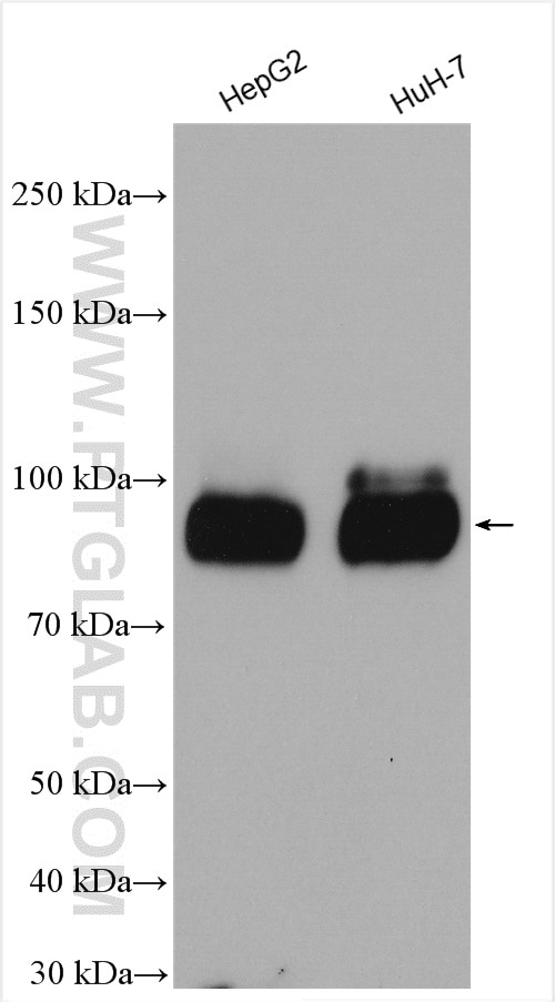
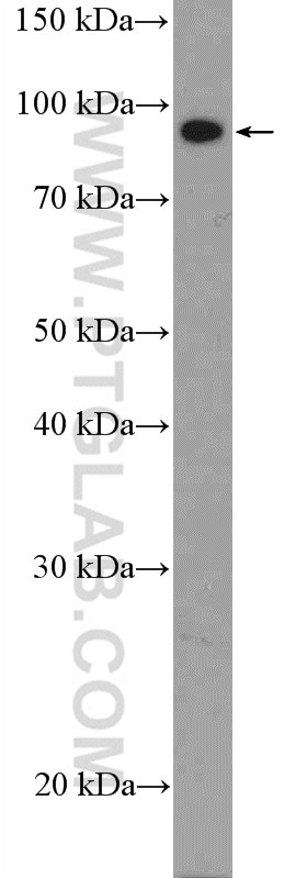
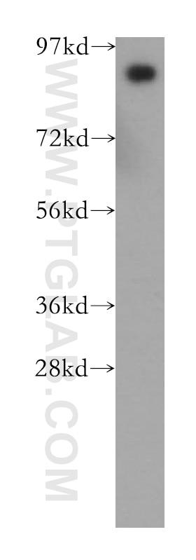
| Résultats positifs en WB | cellules HepG2, cellules HuH-7, cellules MCF-7, tissu hépatique humain |
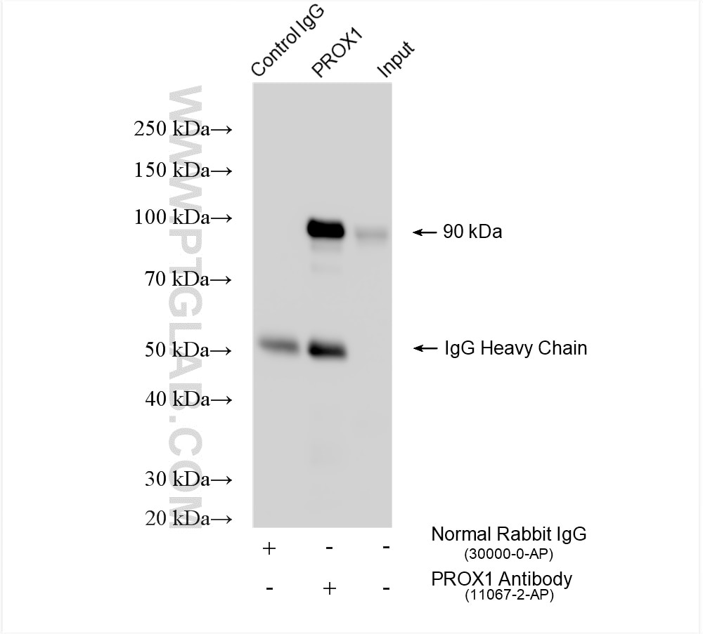
| Résultats positifs en IP | cellules HepG2, |
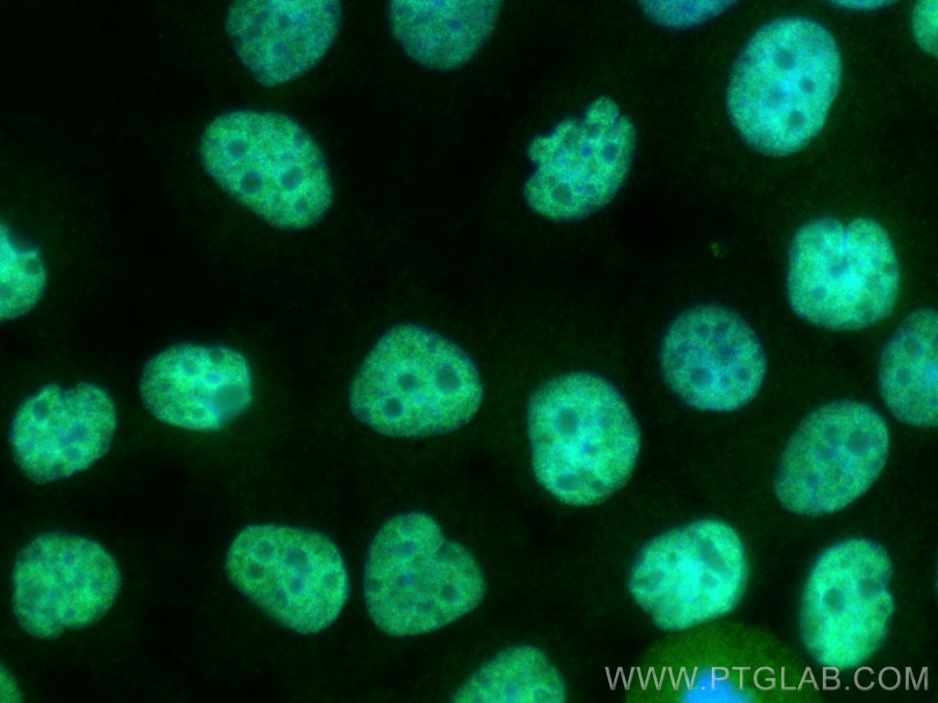
| Résultats positifs en IF/ICC | cellules HuH-7, |
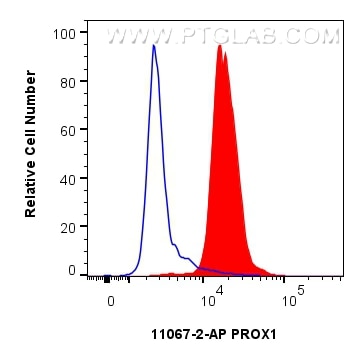
| Résultats positifs en FC (Intra) | cellules HepG2, |
Dilution recommandée
| Application | Dilution |
|---|---|
| Western Blot (WB) | WB : 1:2000-1:10000 |
| Immunoprécipitation (IP) | IP : 0.5-4.0 ug for 1.0-3.0 mg of total protein lysate |
| Immunofluorescence (IF)/ICC | IF/ICC : 1:200-1:800 |
| Flow Cytometry (FC) (INTRA) | FC (INTRA) : 0.25 ug per 10^6 cells in a 100 µl suspension |
| It is recommended that this reagent should be titrated in each testing system to obtain optimal results. | |
| Sample-dependent, check data in validation data gallery | |
Applications publiées
| KD/KO | See 7 publications below |
| WB | See 25 publications below |
| IF | See 19 publications below |
| IP | See 1 publications below |
| ChIP | See 6 publications below |
Informations sur le produit
11067-2-AP cible PROX1 dans les applications de WB, IF/ICC, FC (Intra), IP, ChIP, ELISA et montre une réactivité avec des échantillons Humain
| Réactivité | Humain |
| Réactivité citée | rat, Chèvre, Humain, porc, souris |
| Hôte / Isotype | Lapin / IgG |
| Clonalité | Polyclonal |
| Type | Anticorps |
| Immunogène | PROX1 Protéine recombinante Ag1543 |
| Nom complet | prospero homeobox 1 |
| Masse moléculaire calculée | 737 aa, 83 kDa |
| Poids moléculaire observé | 90 kDa |
| Numéro d’acquisition GenBank | BC024201 |
| Symbole du gène | PROX1 |
| Identification du gène (NCBI) | 5629 |
| Conjugaison | Non conjugué |
| Forme | Liquide |
| Méthode de purification | Purification par affinité contre l'antigène |
| Tampon de stockage | PBS with 0.02% sodium azide and 50% glycerol |
| Conditions de stockage | Stocker à -20°C. Stable pendant un an après l'expédition. L'aliquotage n'est pas nécessaire pour le stockage à -20oC Les 20ul contiennent 0,1% de BSA. |
Informations générales
Prospero-related homeobox 1 (Prox1) is a homeobox transcription factor involved in various developmental processes, including neurogenesis and lymphangiogenesis. In the central nervous system (CNS), it controls the maintenance, proliferation, and differentiation of neural stem cells. This is a rabbit polyclonal antibody raised against part of C-terminal chain of human PROX1. The calcualted molecular weight of PROX1 is 83 kDa.
What is the molecular weight of KEAP1 protein? Is Prox1 post-translationally modified?
The calculated molecular weight of Prox1 protein is 83 kDa but the observed weight is 95 kDa. Prox1 is a subject of SUMO-ylation at K556, which is important for its transcription activity and increases the molecular weight of Prox1 to 110 kDa (PMID: 19706680). Usually there is a pool of modified and unmodified Prox1 protein in cells and therefore it is quite common to observe two specific bands in western blotting using Prox1 antibody.
What is the subcellular localization of Prox1?
Prox1 contains a nuclear localization signal within its N-terminus and resides in the nucleus, where it acts as a transcription factor.
What is the tissue expression pattern of Prox1 in the central nervous system?
Prox1 expression in the central nervous system (CNS) depends on the developmental stage. During embryogenesis Prox1 is present in neuronal precursors located in the subventricular zone, while during fetal development it is additionally present in the cerebral and cerebellar cortex and hippocampus. Postnatally, Prox1 expression in the CNS is restricted to the dentate gyrus of the hippocampus and cerebellum (PMID: 17117441). Therefore, Prox1 can be used as a marker of a subset of neuronal progenitors, as well as a specific marker of dentate gyrus cell lineage, such as type-2 intermediate progenitors and mature granule cells.
Is Prox1 expressed outside of the central nervous system?
Yes. Prox1 is expressed in the developmental stages of the eye lens, liver, pancreas, heart, and the lymphatic system (PMID: 22733308). Prox1 is also expressed post-developmentally in lymphatic endothelial cells and is often used, together with VEGFR-3, as a marker for lymphatic vessels.
Protocole
| Product Specific Protocols | |
|---|---|
| WB protocol for PROX1 antibody 11067-2-AP | Download protocol |
| IF protocol for PROX1 antibody 11067-2-AP | Download protocol |
| IP protocol for PROX1 antibody 11067-2-AP | Download protocol |
| Standard Protocols | |
|---|---|
| Click here to view our Standard Protocols |
Publications
| Species | Application | Title |
|---|---|---|
Cell Metab Neurotensin is an anti-thermogenic peptide produced by lymphatic endothelial cells. | ||
Hepatology Prospero-related homeobox 1 drives angiogenesis of hepatocellular carcinoma through selectively activating interleukin-8 expression.
| ||
Genes Dev The Prox1-Vegfr3 feedback loop maintains the identity and the number of lymphatic endothelial cell progenitors.
| ||
Nat Commun Lipid droplet degradation by autophagy connects mitochondria metabolism to Prox1-driven expression of lymphatic genes and lymphangiogenesis. |
Avis
The reviews below have been submitted by verified Proteintech customers who received an incentive for providing their feedback.
FH Guang (Verified Customer) (11-12-2018) | We use heat antigen retrieval , but we didn't get any result by using this antibody. If it is possible, you can send me another free sample of this type. I will test it by using the enzymatic antigen retrieval or EDTA method to perform the Immunofluorescence.
|
