Anticorps Monoclonal anti-PTK2B
PTK2B Monoclonal Antibody for FC (Intra)
Hôte / Isotype
Mouse / IgG1
Réactivité testée
Humain, porc, rat, souris
Applications
FC (Intra)
Conjugaison
CoraLite® Plus 488 Fluorescent Dye
CloneNo.
2A10G5
N° de cat : CL488-67141
Synonymes
Galerie de données de validation
Applications testées
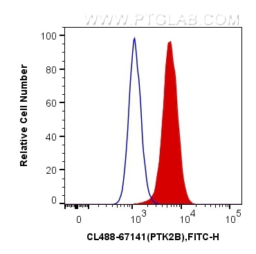
| Résultats positifs en FC (Intra) | cellules Jurkat, |
Dilution recommandée
| Application | Dilution |
|---|---|
| Flow Cytometry (FC) (INTRA) | FC (INTRA) : 0.40 ug per 10^6 cells in a 100 µl suspension |
| It is recommended that this reagent should be titrated in each testing system to obtain optimal results. | |
| Sample-dependent, check data in validation data gallery | |
Informations sur le produit
CL488-67141 cible PTK2B dans les applications de FC (Intra) et montre une réactivité avec des échantillons Humain, porc, rat, souris
| Réactivité | Humain, porc, rat, souris |
| Hôte / Isotype | Mouse / IgG1 |
| Clonalité | Monoclonal |
| Type | Anticorps |
| Immunogène | PTK2B Protéine recombinante Ag11487 |
| Nom complet | PTK2B protein tyrosine kinase 2 beta |
| Masse moléculaire calculée | 1009 aa, 116 kDa |
| Poids moléculaire observé | 112-115 kDa |
| Numéro d’acquisition GenBank | BC042599 |
| Symbole du gène | PTK2B |
| Identification du gène (NCBI) | 2185 |
| Conjugaison | CoraLite® Plus 488 Fluorescent Dye |
| Excitation/Emission maxima wavelengths | 493 nm / 522 nm |
| Forme | Liquide |
| Méthode de purification | Purification par protéine G |
| Tampon de stockage | PBS with 50% glycerol, 0.05% Proclin300, 0.5% BSA |
| Conditions de stockage | Stocker à -20 °C. Éviter toute exposition à la lumière. Stable pendant un an après l'expédition. L'aliquotage n'est pas nécessaire pour le stockage à -20oC Les 20ul contiennent 0,1% de BSA. |
Informations générales
Proline-rich tyrosine kinase 2 (Pyk2; also known as CAK, RAFTK and CADTK) is a cytoplasmic tyrosine kinase implicated to play a role in several intracellular signaling pathways. It is expressed in many cells and tissues and migrated as a 130 kDa band in Western Blotting analysis. The size of PYK2 is 115 kDa, and it expressed in many cells and tissues and migrated as a 130 kDa band, but several lower molecular weight bands (75 kDa, 80 kDa, and 97 kDa) were seen in Western Blotting analysis, possibly due to proteolytic degradation.
Protocole
| Product Specific Protocols | |
|---|---|
| FC protocol for CL Plus 488 PTK2B antibody CL488-67141 | Download protocol |
| Standard Protocols | |
|---|---|
| Click here to view our Standard Protocols |


