- Phare
- Validé par KD/KO
Anticorps Polyclonal de lapin anti-Paxillin
Paxillin Polyclonal Antibody for WB, IHC, IF/ICC, IP, ELISA
Hôte / Isotype
Lapin / IgG
Réactivité testée
Humain, souris et plus (1)
Applications
WB, IHC, IF/ICC, IP, ELISA
Conjugaison
Non conjugué
N° de cat : 10029-1-Ig
Synonymes
Galerie de données de validation
 with sh-Control and sh-Paxillin transfected A431 cells.")
 at dilution of 1:1000 incubated at room temperature for 1.5 hours.")
 at dilution of 1:1000 incubated at room temperature for 1.5 hours.")
 at dilution of 1:1000 incubated at room temperature for 1.5 hours.")
 with A431 cells lysate 1000 ug.")
 at dilution of 1:50 (under 40x lens).")
 at dilution of 1:50 (under 10x lens).")
 at dilution of 1:200 (under 40x lens). Heat mediated antigen retrieval with Tris-EDTA buffer (pH 9.0).")
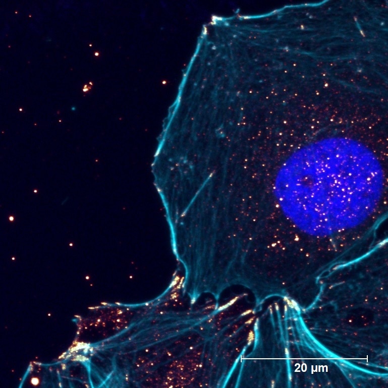
 fixed HUVEC cells using Paxillin antibody (10029-1-Ig) at dilution of 1:200 and CoraLite®488-Conjugated AffiniPure Goat Anti-Rabbit IgG(H+L), CL594-Phalloidin (red). DAPI (blue).")
 at dilution of 1:50 and Alexa Fluor 488-conjugated AffiniPure Goat Anti-Rabbit IgG(H+L). The cytoskeleton was labelled in red with 66031-1-Ig (alpha tubulin). DAPI (blue).")
 fixed HepG2 cells using 10029-1-Ig (Paxillin antibody) at dilution of 1:50 and Alexa Fluor 488-conjugated AffiniPure Goat Anti-Rabbit IgG(H+L).")
 fixed NIH/3T3 cells using 10029-1-Ig(Paxillin antibody) at dilution of 1:50 and Alexa Fluor 488-conjugated AffiniPure Goat Anti-Rabbit IgG(H+L).")
Applications testées
| Résultats positifs en WB | cellules COLO 320, cellules A431, cellules Jurkat, cellules MCF-7 |
| Résultats positifs en IP | cellules A431, |
| Résultats positifs en IHC | tissu de cancer du foie humain, il est suggéré de démasquer l'antigène avec un tampon de TE buffer pH 9.0; (*) À défaut, 'le démasquage de l'antigène peut être 'effectué avec un tampon citrate pH 6,0. |
| Résultats positifs en IF/ICC | cellules HUVEC, cellules HepG2, cellules NIH/3T3 |
Dilution recommandée
| Application | Dilution |
|---|---|
| Western Blot (WB) | WB : 1:500-1:2000 |
| Immunoprécipitation (IP) | IP : 0.5-4.0 ug for 1.0-3.0 mg of total protein lysate |
| Immunohistochimie (IHC) | IHC : 1:50-1:500 |
| Immunofluorescence (IF)/ICC | IF/ICC : 1:50-1:500 |
| It is recommended that this reagent should be titrated in each testing system to obtain optimal results. | |
| Sample-dependent, check data in validation data gallery | |
Applications publiées
| WB | See 11 publications below |
| IHC | See 3 publications below |
| IF | See 9 publications below |
Informations sur le produit
10029-1-Ig cible Paxillin dans les applications de WB, IHC, IF/ICC, IP, ELISA et montre une réactivité avec des échantillons Humain, souris
| Réactivité | Humain, souris |
| Réactivité citée | rat, Humain, souris |
| Hôte / Isotype | Lapin / IgG |
| Clonalité | Polyclonal |
| Type | Anticorps |
| Immunogène | Peptide |
| Nom complet | paxillin |
| Masse moléculaire calculée | 68 kDa |
| Poids moléculaire observé | 68 kDa |
| Numéro d’acquisition GenBank | NM_002859 |
| Symbole du gène | Paxillin |
| Identification du gène (NCBI) | 5829 |
| Conjugaison | Non conjugué |
| Forme | Liquide |
| Méthode de purification | Purification par protéine A |
| Tampon de stockage | PBS with 0.02% sodium azide and 50% glycerol |
| Conditions de stockage | Stocker à -20°C. Stable pendant un an après l'expédition. L'aliquotage n'est pas nécessaire pour le stockage à -20oC Les 20ul contiennent 0,1% de BSA. |
Informations générales
PXN (paxillin) is a 68 kDa scaffold protein that interacts with multiple structural and signaling proteins and regulates cell adhesion, migration, proliferation, and apoptosis. PXN is thought to play an important role in tumor migration, invasion, and metastasis (21045234). PXN has been identified as a direct substrate of protein tyrosine phosphatase receptor-type T (PTPRT), a potent tumor suppressor gene. Increased phospho-PXN at tyrosine residue 88 (Y88) has been found as a common feature of human colon cancers (20133777).
Protocole
| Product Specific Protocols | |
|---|---|
| WB protocol for Paxillin antibody 10029-1-Ig | Download protocol |
| IHC protocol for Paxillin antibody 10029-1-Ig | Download protocol |
| IF protocol for Paxillin antibody 10029-1-Ig | Download protocol |
| IP protocol for Paxillin antibody 10029-1-Ig | Download protocol |
| Standard Protocols | |
|---|---|
| Click here to view our Standard Protocols |
Publications
| Species | Application | Title |
|---|---|---|
Mol Cell Direct epitranscriptomic regulation of mammalian translation initiation through N4-acetylcytidine. | ||
Sci Adv Bioactive fiber-reinforced hydrogel to tailor cell microenvironment for structural and functional regeneration of myotendinous junction | ||
Bone Res Impairment of rigidity sensing caused by mutant TP53 gain of function in osteosarcoma | ||
Cell Rep Mechanotransduction in response to ECM stiffening impairs cGAS immune signaling in tumor cells | ||
Cell Death Dis ZC3H15 promotes glioblastoma progression through regulating EGFR stability. | ||
Acta Pharmacol Sin Reduced intracellular chloride concentration impairs angiogenesis by inhibiting oxidative stress-mediated VEGFR2 activation. |
Avis
The reviews below have been submitted by verified Proteintech customers who received an incentive for providing their feedback.
FH Thomas (Verified Customer) (09-22-2022) | Works in WB (pulldown via PAK1) and IF (costained with F-Actin in cyan)
 |
FH Boyan (Verified Customer) (03-11-2019) | For WB, besides the expected band, it also recognised some other non-specific bands; for IF, it could label the cell cortex localisation, partially co-localised with Actin.
|

