Anticorps Polyclonal de lapin anti-RLBP1
RLBP1 Polyclonal Antibody for WB, IHC, IF/ICC, IF-P, ELISA
Hôte / Isotype
Lapin / IgG
Réactivité testée
Humain, rat, souris et plus (1)
Applications
WB, IHC, IF/ICC, IF-P, ELISA
Conjugaison
Non conjugué
N° de cat : 15356-1-AP
Synonymes
Galerie de données de validation
 at dilution of 1:1000 incubated at room temperature for 1.5 hours.")
 at dilution of 1:2000 incubated at room temperature for 1.5 hours.")
 at dilution of 1:500 incubated at room temperature for 1.5 hours.")
 at dilution of 1:50 (under 10x lens).")
 at dilution of 1:50 (under 40x lens).")
 at dilution of 1:200 (under 10x lens). Heat mediated antigen retrieval with Tris-EDTA buffer (pH 9.0).")
 at dilution of 1:200 (under 40x lens). Heat mediated antigen retrieval with Tris-EDTA buffer (pH 9.0).")
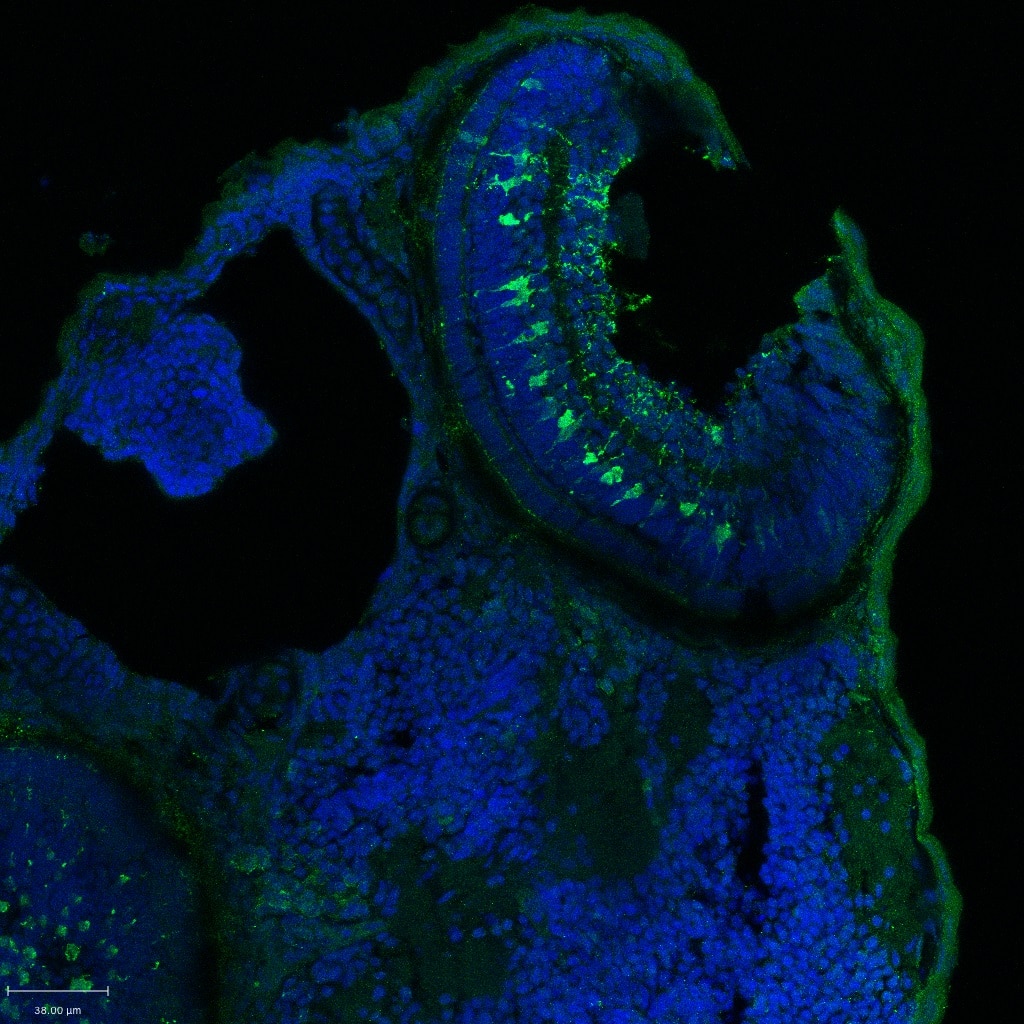
 fixed paraffin-embedded mouse eye tissue using RLBP1 antibody (15356-1-AP) at dilution of 1:400 and CoraLite®488-Conjugated AffiniPure Goat Anti-Rabbit IgG(H+L) (SA00013-2). Heat mediated antigen retrieval with Tris-EDTA buffer (pH 9.0).")
 fixed paraffin-embedded mouse eye tissue using RLBP1 antibody (15356-1-AP) at dilution of 1:400 and CoraLite®488-Conjugated AffiniPure Goat Anti-Rabbit IgG(H+L) (SA00013-2). Heat mediated antigen retrieval with Tris-EDTA buffer (pH 9.0).")
 fixed HeLa cells using RLBP1 antibody (15356-1-AP) at dilution of 1:200 and CoraLite®488-Conjugated AffiniPure Goat Anti-Rabbit IgG(H+L).")
 fixed Y79 cells using RLBP1 antibody (15356-1-AP) at dilution of 1:200 and CoraLite®488-Conjugated AffiniPure Goat Anti-Rabbit IgG(H+L).")
 generated from human induced pluripotent stem cells (iPSCs) and fixed with 4% PFA. Stained for Tubulin beta 3/TUJ1 using 66375-1-Ig at 1:500 dilution (green) and CRALBP using 15356-1-AP at 1:400 (red). Nuclear stain DAPI (blue). Scale bar = 50 µm. Data generated by Alessandro Bellapianta at Johannes Kepler Universitat, Austria.")
Applications testées
| Résultats positifs en WB | tissu oculaire de souris, cellules HepG2, tissu oculaire de rat, tissu rétinien de souris |
| Résultats positifs en IHC | tissu oculaire de souris, tissu oculaire humain il est suggéré de démasquer l'antigène avec un tampon de TE buffer pH 9.0; (*) À défaut, 'le démasquage de l'antigène peut être 'effectué avec un tampon citrate pH 6,0. |
| Résultats positifs en IF-P | tissu oculaire de souris, Retinal organoids |
| Résultats positifs en IF/ICC | cellules HeLa, cellules Y79 |
Dilution recommandée
| Application | Dilution |
|---|---|
| Western Blot (WB) | WB : 1:1000-1:4000 |
| Immunohistochimie (IHC) | IHC : 1:50-1:500 |
| Immunofluorescence (IF)-P | IF-P : 1:200-1:800 |
| Immunofluorescence (IF)/ICC | IF/ICC : 1:50-1:500 |
| It is recommended that this reagent should be titrated in each testing system to obtain optimal results. | |
| Sample-dependent, check data in validation data gallery | |
Applications publiées
| WB | See 5 publications below |
| IHC | See 2 publications below |
| IF | See 13 publications below |
Informations sur le produit
15356-1-AP cible RLBP1 dans les applications de WB, IHC, IF/ICC, IF-P, ELISA et montre une réactivité avec des échantillons Humain, rat, souris
| Réactivité | Humain, rat, souris |
| Réactivité citée | rat, Humain, poisson-zèbre, souris |
| Hôte / Isotype | Lapin / IgG |
| Clonalité | Polyclonal |
| Type | Anticorps |
| Immunogène | RLBP1 Protéine recombinante Ag7602 |
| Nom complet | retinaldehyde binding protein 1 |
| Masse moléculaire calculée | 36 kDa |
| Poids moléculaire observé | 36 kDa |
| Numéro d’acquisition GenBank | BC004199 |
| Symbole du gène | RLBP1 |
| Identification du gène (NCBI) | 6017 |
| Conjugaison | Non conjugué |
| Forme | Liquide |
| Méthode de purification | Purification par affinité contre l'antigène |
| Tampon de stockage | PBS with 0.02% sodium azide and 50% glycerol |
| Conditions de stockage | Stocker à -20°C. Stable pendant un an après l'expédition. L'aliquotage n'est pas nécessaire pour le stockage à -20oC Les 20ul contiennent 0,1% de BSA. |
Informations générales
RLBP1 (Retinaldehyde-binding protein 1) is also named as cellular retinaldehyde-binding protein (CRALBP). CRALBP, a 36-kDa aqueous soluble carrier, is expressed in abundance by both retinal pigment epithelial (RPE) and Muller cells of retina and is important for rod- and cone-driven vision (PMID: 32188692). Retinal RLBP1 expression was decreased in diabetes, and its overexpression in Müller glia mitigated DR-associated neurovascular degeneration (PMID: 33674409).
Protocole
| Product Specific Protocols | |
|---|---|
| WB protocol for RLBP1 antibody 15356-1-AP | Download protocol |
| IHC protocol for RLBP1 antibody 15356-1-AP | Download protocol |
| IF protocol for RLBP1 antibody 15356-1-AP | Download protocol |
| Standard Protocols | |
|---|---|
| Click here to view our Standard Protocols |
Publications
| Species | Application | Title |
|---|---|---|
Aging Cell Müller Glia maintain their regenerative potential despite degeneration in the aged zebrafish retina. | ||
Mol Vis Morphological, biochemical, and transcriptomic characterization of iPSC-derived human RPE cells from normal and Smith-Lemli-Opitz syndrome patients | ||
Diabetes CD40 in Retinal Müller Cells Induces P2X7-Dependent Cytokine Expression in Macrophages/Microglia in Diabetic Mice and Development of Early Experimental Diabetic Retinopathy. | ||
FASEB J A cell-penetrating CD40-TRAF2,3 blocking peptide diminishes inflammation and neuronal loss after ischemia/reperfusion. | ||
iScience Induced retinal pigment epithelial cells with anti-epithelial-to-mesenchymal transition ability delay retinal degeneration | ||
Invest Ophthalmol Vis Sci CD40 Upregulation in the Retina of Patients With Diabetic Retinopathy: Association With TRAF2/TRAF6 Upregulation and Inflammatory Molecule Expression |
Avis
The reviews below have been submitted by verified Proteintech customers who received an incentive for providing their feedback.
FH Ryan (Verified Customer) (01-24-2018) | Antigen retrieval in NaCit 0.1M ph 6
 |

