- Phare
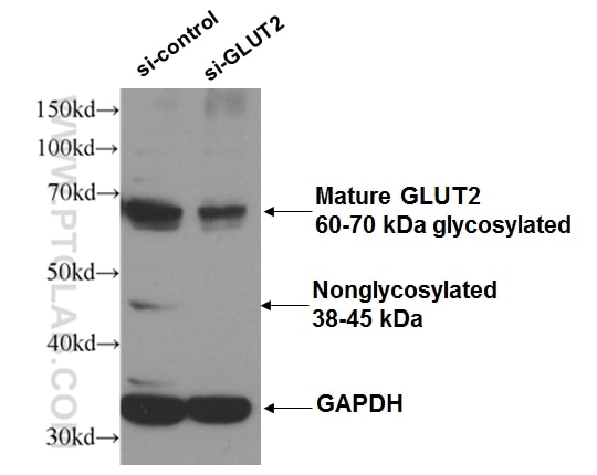
- Validé par KD/KO
Anticorps Polyclonal de lapin anti-GLUT2
GLUT2 Polyclonal Antibody for WB, IHC, IP, ELISA
Hôte / Isotype
Lapin / IgG
Réactivité testée
Humain, rat, souris et plus (2)
Applications
WB, IHC, IF, IP, ELISA
Conjugaison
Non conjugué
N° de cat : 20436-1-AP
Synonymes
Galerie de données de validation
Applications testées
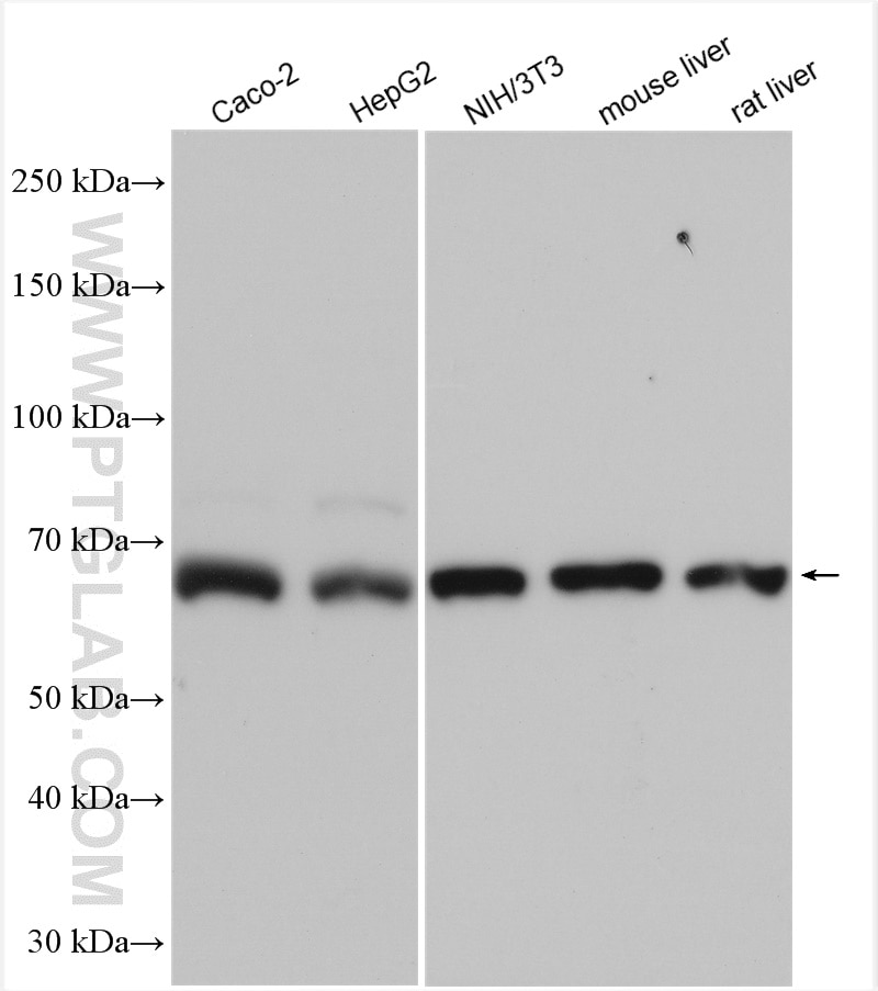
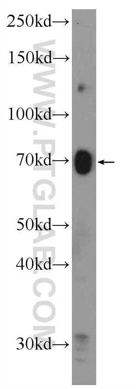
| Résultats positifs en WB | cellules Caco-2, cellules HEK 293, cellules HepG2, cellules NIH/3T3, tissu hépatique de rat, tissu hépatique de souris, tissu rénal de souris |
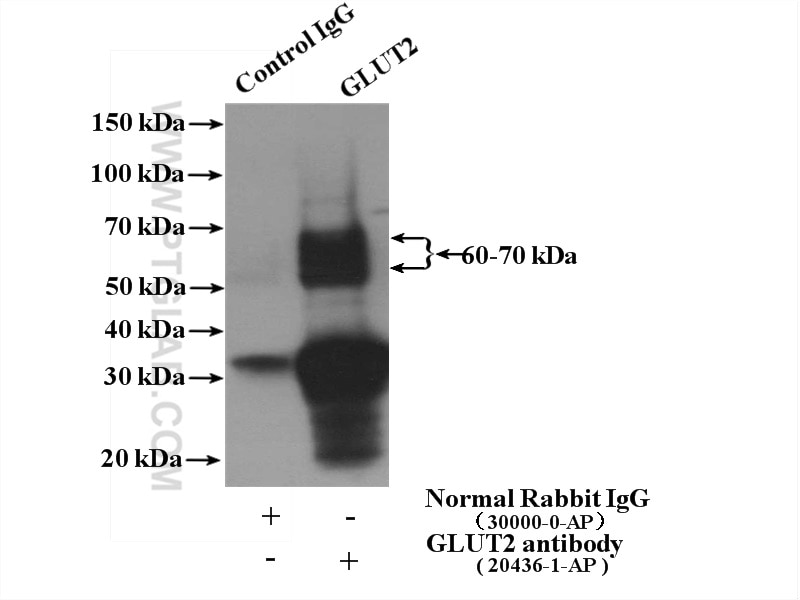
| Résultats positifs en IP | tissu rénal de souris |
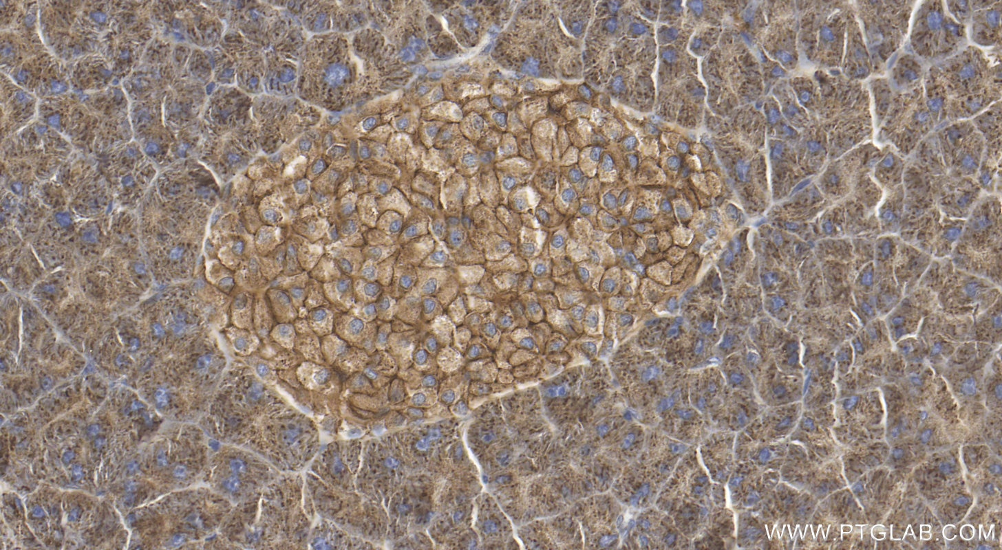
| Résultats positifs en IHC | tissu pancréatique de souris, il est suggéré de démasquer l'antigène avec un tampon de TE buffer pH 9.0; (*) À défaut, 'le démasquage de l'antigène peut être 'effectué avec un tampon citrate pH 6,0. |
Dilution recommandée
| Application | Dilution |
|---|---|
| Western Blot (WB) | WB : 1:500-1:3000 |
| Immunoprécipitation (IP) | IP : 0.5-4.0 ug for 1.0-3.0 mg of total protein lysate |
| Immunohistochimie (IHC) | IHC : 1:500-1:2000 |
| It is recommended that this reagent should be titrated in each testing system to obtain optimal results. | |
| Sample-dependent, check data in validation data gallery | |
Applications publiées
| WB | See 71 publications below |
| IHC | See 15 publications below |
| IF | See 13 publications below |
Informations sur le produit
20436-1-AP cible GLUT2 dans les applications de WB, IHC, IF, IP, ELISA et montre une réactivité avec des échantillons Humain, rat, souris
| Réactivité | Humain, rat, souris |
| Réactivité citée | rat, Humain, porc, souris, Lasiopodomys Brandtii |
| Hôte / Isotype | Lapin / IgG |
| Clonalité | Polyclonal |
| Type | Anticorps |
| Immunogène | GLUT2 Protéine recombinante Ag14204 |
| Nom complet | solute carrier family 2 (facilitated glucose transporter), member 2 |
| Masse moléculaire calculée | 57 kDa |
| Poids moléculaire observé | 60-70 kDa, 38-45 kDa |
| Numéro d’acquisition GenBank | BC060041 |
| Symbole du gène | GLUT2 |
| Identification du gène (NCBI) | 6514 |
| Conjugaison | Non conjugué |
| Forme | Liquide |
| Méthode de purification | Purification par affinité contre l'antigène |
| Tampon de stockage | PBS with 0.02% sodium azide and 50% glycerol |
| Conditions de stockage | Stocker à -20°C. Stable pendant un an après l'expédition. L'aliquotage n'est pas nécessaire pour le stockage à -20oC Les 20ul contiennent 0,1% de BSA. |
Informations générales
Glucose transporter 2 (GLUT2), also known as solute carrier family 2, facilitated glucose transporter member 2 (SLC2A2), is a transporter protein regulating glucose transport across cell membranes in an insulin-independent manner.
What is the molecular weight of GLUT2? Is GLUT2 post-translationally modified?
GLUT2 can be N-glycosylated. The molecular weight of mature glycosylated GLUT2 transporter is between 55 and 65 kDa, but a minor 38-kDa form is often detected in hepatocytes that may represent a non-glycosylated or proteolytic product (PMID: 12101013).
What is the subcellular localization of GLUT2?
Glucose transporters, including GLUT2, are multiple-pass integral membrane proteins. GLUT2 is present at the plasma membrane but has also been reported to be present at intracellular membranes, including the endoplasmic reticulum (PMID: 12101013).
What molecules can be transported by GLUT2?
Although the main substrate of GLUT2 transport is glucose, it can also transport galactose, mannose, and fructose.
What is the tissue expression pattern of GLUT2?
GLUT2 is expressed in many cell types. High expression of GLUT2 is observed in hepatocytes, where GLUT2 mediates glucose uptake and release regulating glucose levels in blood, in intestinal cells, where it regulates glucose transport at the basolateral membrane and to a lesser extent from the apical membrane, and in the kidney proximal tubule, where it is involved in glucose reabsorption and is present at the basolateral membrane.
Protocole
| Product Specific Protocols | |
|---|---|
| WB protocol for GLUT2 antibody 20436-1-AP | Download protocol |
| IHC protocol for GLUT2 antibody 20436-1-AP | Download protocol |
| IP protocol for GLUT2 antibody 20436-1-AP | Download protocol |
| Standard Protocols | |
|---|---|
| Click here to view our Standard Protocols |
Publications
| Species | Application | Title |
|---|---|---|
Clin Transl Med USP7 promotes non-small-cell lung cancer cell glycolysis and survival by stabilizing and activating c-Abl | ||
Cell Mol Gastroenterol Hepatol Patient-Derived Mutant Forms of NFE2L2/NRF2 Drive Aggressive Murine Hepatoblastomas. | ||
Mol Metab T1R2 receptor-mediated glucose sensing in the upper intestine potentiates glucose absorption through activation of local regulatory pathways. | ||
Metabolism Rheb1 promotes glucose-stimulated insulin secretion in human and mouse β-cells by upregulating GLUT expression. |
Avis
The reviews below have been submitted by verified Proteintech customers who received an incentive for providing their feedback.
FH Vignesh (Verified Customer) (09-15-2025) | results are excellent
|
FH Balawant (Verified Customer) (07-25-2022) | This antibody I used and it is working great. I will recommend this antibody for Immunoblting.
|
FH Susmita (Verified Customer) (04-13-2022) | This antibody is working nicely for my WB
|
FH Boyan (Verified Customer) (08-24-2021) | Not conclusive. This antibody primarily recognized a band at ~ 90 kd, which did not respond to a Glut2 siRNA treatment. It also weakly recognized a band at ~ 70 kd, which responded to de-glycosylation treatment.
|
FH Iram (Verified Customer) (09-17-2020) | Excellent GLUT-2 Antibody. Giving a very good results for WB and IF.
|
FH Pradeep (Verified Customer) (09-17-2020) | Very good antibody for western blot. I checked in INS-1 cell line.
|
