- Phare
- Validé par KD/KO
Anticorps Polyclonal de lapin anti-GLUT3
GLUT3 Polyclonal Antibody for WB, IHC, IF/ICC, ELISA
Hôte / Isotype
Lapin / IgG
Réactivité testée
Humain, rat, souris et plus (1)
Applications
WB, IHC, IF/ICC, IP, ELISA
Conjugaison
Non conjugué
N° de cat : 20403-1-AP
Synonymes
Galerie de données de validation
 at dilution of 1:1500 incubated at room temperature for 1.5 hours.")
 at dilution of 1:5000 incubated at room temperature for 1.5 hours.")
 at dilution of 1:500 incubated at room temperature for 1.5 hours.")
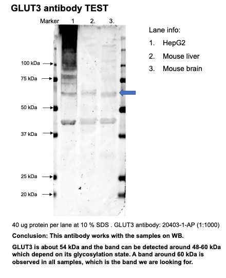
 at dilution of 1:1000 incubated at room temperature for 1.5 hours.")
 at dilution of 1:4000 (under 10x lens). Heat mediated antigen retrieval with Tris-EDTA buffer (pH 9.0).")
 at dilution of 1:4000 (under 40x lens). Heat mediated antigen retrieval with Tris-EDTA buffer (pH 9.0).")
 at dilution of 1:1000 (under 40x lens). Heat mediated antigen retrieval with Tris-EDTA buffer (pH 9.0).")
 at dilution of 1:1000 (under 10x lens). Heat mediated antigen retrieval with Tris-EDTA buffer (pH 9.0).")
 at dilution of 1:1000 (under 40x lens). Heat mediated antigen retrieval with Tris-EDTA buffer (pH 9.0).")
 at dilution of 1:1000 (under 10x lens). Heat mediated antigen retrieval with Tris-EDTA buffer (pH 9.0).")
. Heat mediated antigen retrieval with Tris-EDTA buffer (pH 9.0).")
. Heat mediated antigen retrieval with Tris-EDTA buffer (pH 9.0).")
 at dilution of 1:200 (under 10x lens). Heat mediated antigen retrieval with Tris-EDTA buffer (pH 9.0).")
 at dilution of 1:200 (under 40x lens). Heat mediated antigen retrieval with Tris-EDTA buffer (pH 9.0).")
 at dilution of 1:800 (under 10x lens). Heat mediated antigen retrieval with Tris-EDTA buffer (pH 9.0).")
 at dilution of 1:800 (under 40x lens). Heat mediated antigen retrieval with Tris-EDTA buffer (pH 9.0).")
 fixed Caco-2 cells using GLUT3 antibody (20403-1-AP) at dilution of 1:200 and CoraLite®488-Conjugated AffiniPure Goat Anti-Rabbit IgG(H+L).")
 fixed Caco-2 cells using GLUT3 antibody (20403-1-AP) at dilution of 1:400 and CoraLite®488-Conjugated AffiniPure Goat Anti-Rabbit IgG(H+L).")
Applications testées
| Résultats positifs en WB | cellules HEK-293, cellules C6, cellules HeLa, cellules HepG2, cellules Jurkat, cellules U-251, tissu cérébral de rat, tissu cérébral de souris |
| Résultats positifs en IHC | tissu testiculaire de souris, tissu cérébral de souris, tissu de cancer du poumon humain, tissu de cancer du sein humain, tissu placentaire humain il est suggéré de démasquer l'antigène avec un tampon de TE buffer pH 9.0; (*) À défaut, 'le démasquage de l'antigène peut être 'effectué avec un tampon citrate pH 6,0. |
| Résultats positifs en IF/ICC | cellules Caco-2, |
Dilution recommandée
| Application | Dilution |
|---|---|
| Western Blot (WB) | WB : 1:2000-1:10000 |
| Immunohistochimie (IHC) | IHC : 1:2000-1:8000 |
| Immunofluorescence (IF)/ICC | IF/ICC : 1:200-1:800 |
| It is recommended that this reagent should be titrated in each testing system to obtain optimal results. | |
| Sample-dependent, check data in validation data gallery | |
Applications publiées
| KD/KO | See 1 publications below |
| WB | See 67 publications below |
| IHC | See 12 publications below |
| IF | See 18 publications below |
| IP | See 1 publications below |
Informations sur le produit
20403-1-AP cible GLUT3 dans les applications de WB, IHC, IF/ICC, IP, ELISA et montre une réactivité avec des échantillons Humain, rat, souris
| Réactivité | Humain, rat, souris |
| Réactivité citée | rat, Chèvre, Humain, souris |
| Hôte / Isotype | Lapin / IgG |
| Clonalité | Polyclonal |
| Type | Anticorps |
| Immunogène | GLUT3 Protéine recombinante Ag14203 |
| Nom complet | solute carrier family 2 (facilitated glucose transporter), member 3 |
| Masse moléculaire calculée | 496 aa, 54 kDa |
| Poids moléculaire observé | 54-60 kDa |
| Numéro d’acquisition GenBank | BC039196 |
| Symbole du gène | GLUT3 |
| Identification du gène (NCBI) | 6515 |
| Conjugaison | Non conjugué |
| Forme | Liquide |
| Méthode de purification | Purification par affinité contre l'antigène |
| Tampon de stockage | PBS with 0.02% sodium azide and 50% glycerol |
| Conditions de stockage | Stocker à -20°C. Stable pendant un an après l'expédition. L'aliquotage n'est pas nécessaire pour le stockage à -20oC Les 20ul contiennent 0,1% de BSA. |
Informations générales
Glucose transporter 3 (GLUT3), also known as solute carrier family 2, facilitated glucose transporter member 3 (SLC2A3), is a transporter protein regulating glucose transport across cell membranes and is a primary glucose transporter in neurons.
What is the molecular weight of GLUT3? Is GLUT3 post-translationally modified?
The molecular weight of GLUT3 transporter is 49-52 kDa and depends on the glycosylation pattern, which is tissue-specific (PMID: 9253355 and 9889355). GLUT3 can be N-glycosylated.
What is the subcellular localization of GLUT3?
Glucose transporters, including GLUT3, are multiple-pass integral membrane proteins. GLUT3 is present at the plasma membrane but is also a subject of recycling between the plasma membrane and endosomes.
What molecules can be transported by GLUT3?
Although the main substrate of GLUT3 transport is glucose, it can also transport mannose, galactose, and xylose.
What is the tissue expression pattern of GLUT3?
GLUT1 and GLUT3 are important for the transport of glucose into the central nervous system. While GLUT1 transports glucose through blood vessels and into astrocytes, GLUT3 is predominantly expressed in neurons, being their main glucose transporter (PMID: 9302083), and in testis (PMID: 18577699).
Protocole
| Product Specific Protocols | |
|---|---|
| WB protocol for GLUT3 antibody 20403-1-AP | Download protocol |
| IHC protocol for GLUT3 antibody 20403-1-AP | Download protocol |
| IF protocol for GLUT3 antibody 20403-1-AP | Download protocol |
| Standard Protocols | |
|---|---|
| Click here to view our Standard Protocols |
Publications
| Species | Application | Title |
|---|---|---|
Cell Metab Acetate enables metabolic fitness and cognitive performance during sleep disruption | ||
Nat Aging Activation of AMPK by GLP-1R agonists mitigates Alzheimer-related phenotypes in transgenic mice | ||
Neuron Sympathetic nerve-enteroendocrine L cell communication modulates GLP-1 release, brain glucose utilization, and cognitive function | ||
Mol Cancer CircEZH2/miR-133b/IGF2BP2 aggravates colorectal cancer progression via enhancing the stability of m6A-modified CREB1 mRNA. | ||
Cell Death Differ A viral interferon regulatory factor degrades RNA-binding protein hnRNP Q1 to enhance aerobic glycolysis via recruiting E3 ubiquitin ligase KLHL3 and decaying GDPD1 mRNA. | ||
Autophagy Newcastle disease virus degrades SIRT3 via PINK1-PRKN-dependent mitophagy to reprogram energy metabolism in infected cells. |
Avis
The reviews below have been submitted by verified Proteintech customers who received an incentive for providing their feedback.
FH balawant (Verified Customer) (12-25-2023) | This antibody is very specific and has no background.
|
FH Hua (Verified Customer) (02-03-2023) | This antibody works ok. There is one strong non-specific band around 40 kDa.
 |

