- Phare
- Validé par KD/KO
Anticorps Polyclonal de lapin anti-ST6GAL1
ST6GAL1 Polyclonal Antibody for WB, IHC, IP, ELISA
Hôte / Isotype
Lapin / IgG
Réactivité testée
Humain, souris et plus (1)
Applications
WB, IHC, IF, IP, ELISA
Conjugaison
Non conjugué
N° de cat : 14355-1-AP
Synonymes
Galerie de données de validation
 with sh-Control and sh-ST6GAL1 transfected HepG2 cells.")
 at dilution of 1:3000 incubated at room temperature for 1.5 hours.")
 at dilution of 1:600 incubated at room temperature for 1.5 hours.")
 at dilution of 1:600 incubated at room temperature for 1.5 hours.")
 at dilution of 1:600 incubated at room temperature for 1.5 hours.")
 at dilution of 1:1500 incubated at room temperature for 1.5 hours.")
 with Raji cells lysate 1800 ug.")
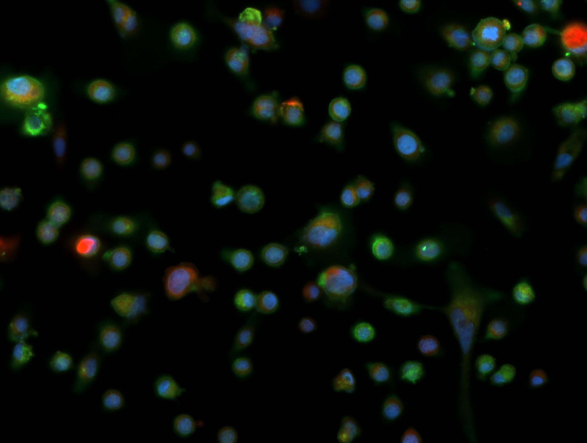
 at dilution of 1:600 (under 20x lens). Heat mediated antigen retrieval with Tris-EDTA buffer (pH 9.0).")
 at dilution of 1:600 (under 20x lens). Heat mediated antigen retrieval with Tris-EDTA buffer (pH 9.0).")
 at dilution of 1:200 (under 10x lens).")
 at dilution of 1:200 (under 40x lens).")
 at dilution of 1:200 (under 20x lens). Heat mediated antigen retrieval with Tris-EDTA buffer (pH 9.0).")
 at dilution of 1:200 (under 20x lens). Heat mediated antigen retrieval with Tris-EDTA buffer (pH 9.0).")
Applications testées
| Résultats positifs en WB | cellules HepG2, cellules HuH-7, cellules Jurkat, cellules L02, cellules U-937, tissu splénique de souris |
| Résultats positifs en IP | cellules Raji, |
| Résultats positifs en IHC | tissu de cancer du côlon humain, tissu cutané humain il est suggéré de démasquer l'antigène avec un tampon de TE buffer pH 9.0; (*) À défaut, 'le démasquage de l'antigène peut être 'effectué avec un tampon citrate pH 6,0. |
Dilution recommandée
| Application | Dilution |
|---|---|
| Western Blot (WB) | WB : 1:1000-1:6000 |
| Immunoprécipitation (IP) | IP : 0.5-4.0 ug for 1.0-3.0 mg of total protein lysate |
| Immunohistochimie (IHC) | IHC : 1:300-1:1200 |
| It is recommended that this reagent should be titrated in each testing system to obtain optimal results. | |
| Sample-dependent, check data in validation data gallery | |
Applications publiées
| KD/KO | See 4 publications below |
| WB | See 9 publications below |
| IHC | See 6 publications below |
| IF | See 3 publications below |
Informations sur le produit
14355-1-AP cible ST6GAL1 dans les applications de WB, IHC, IF, IP, ELISA et montre une réactivité avec des échantillons Humain, souris
| Réactivité | Humain, souris |
| Réactivité citée | rat, Humain, souris |
| Hôte / Isotype | Lapin / IgG |
| Clonalité | Polyclonal |
| Type | Anticorps |
| Immunogène | ST6GAL1 Protéine recombinante Ag5705 |
| Nom complet | ST6 beta-galactosamide alpha-2,6-sialyltranferase 1 |
| Masse moléculaire calculée | 47 kDa |
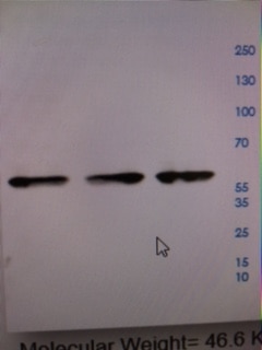
| Poids moléculaire observé | 43-45 kDa, 50-70 kDa |
| Numéro d’acquisition GenBank | BC040009 |
| Symbole du gène | ST6GAL1 |
| Identification du gène (NCBI) | 6480 |
| Conjugaison | Non conjugué |
| Forme | Liquide |
| Méthode de purification | Purification par affinité contre l'antigène |
| Tampon de stockage | PBS with 0.02% sodium azide and 50% glycerol |
| Conditions de stockage | Stocker à -20°C. Stable pendant un an après l'expédition. L'aliquotage n'est pas nécessaire pour le stockage à -20oC Les 20ul contiennent 0,1% de BSA. |
Informations générales
ST6GAL1 (β-galactoside α-2-6 sialyl transferase1; also known as ST6N or CD75) is a sialyltransferase mediating the glycosylation of proteins and lipids to form functionally important glycoproteins and glycolipids in the Golgi compartment. It is principally expressed in liver, placenta, and skeletal muscle. ST6GAL1 undergoes proteolytic process to generate soluble form from membrane form. Western blot analysis of human liver using this antibody detects both isoforms between 43-50 kDa. Higher molecular weight of bands around 50-70 kDa can also be observed with glycosylation modification. (PMID: 15049997, 23358684)
Protocole
| Product Specific Protocols | |
|---|---|
| WB protocol for ST6GAL1 antibody 14355-1-AP | Download protocol |
| IHC protocol for ST6GAL1 antibody 14355-1-AP | Download protocol |
| IP protocol for ST6GAL1 antibody 14355-1-AP | Download protocol |
| Standard Protocols | |
|---|---|
| Click here to view our Standard Protocols |
Publications
| Species | Application | Title |
|---|---|---|
Immunity Immune checkpoint therapy-elicited sialylation of IgG antibodies impairs antitumorigenic type I interferon responses in hepatocellular carcinoma | ||
Int J Cancer Modification of α2,6-sialylation mediates the invasiveness and tumorigenicity of non-small cell lung cancer cells in vitro and in vivo via Notch1/Hes1/MMPs pathway.
| ||
Mol Cell Proteomics Integrated Systems Analysis of the Murine and Human Pancreatic Cancer Glycomes Reveals a Tumor-Promoting Role for ST6GAL1
| ||
Front Oncol High-Risk HPV16 E6 Activates the cGMP/PKG Pathway Through Glycosyltransferase ST6GAL1 in Cervical Cancer Cells.
| ||
Am J Cancer Res α1,6-Fucosyltransferase (FUT8) regulates the cancer-promoting capacity of cancer-associated fibroblasts (CAFs) by modifying EGFR core fucosylation (CF) in non-small cell lung cancer (NSCLC). | ||
Int J Food Sci Nutr Ellagic acid alleviates TNBS-induced intestinal barrier dysfunction by regulating mucin secretion and maintaining tight junction integrity in rats |
Avis
The reviews below have been submitted by verified Proteintech customers who received an incentive for providing their feedback.
FH Boyan (Verified Customer) (04-23-2024) | The strong band around 60 kd is a non-specific band. 1) This band doesn't respond to RNAi (I have another St6gal1 antibody which can confirm the RNAi was working), and 2) this band doesn't respond to PNGase deglycosylation (St6gal1 carries glycosylation, which should show a downshift after PNGase treatment).
|
FH Vanessa (Verified Customer) (01-12-2024) | 1:1000 dilution of the primary antibody accompanied by the recommended blocking, washing and incubation steps resulted in crisp bands with no background.
|
FH Maggie (Verified Customer) (11-15-2023) | Overnight incubation
 |
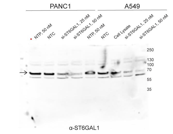
FH Faezeh (Verified Customer) (01-29-2023) | It showed two bands in A549 and PANC1 cell lines, we validated that top band is the correct one using siRNA against ST6GAL1.
 |
FH Dawn (Verified Customer) (02-22-2022) | It is difficult to find good antibodies for glycoslytransferase genes, but this is a good one. It works in multiple cell lines as well.
 |
FH Patricia (Verified Customer) (02-25-2020) | Did not work- even for positive control
|

