- Phare
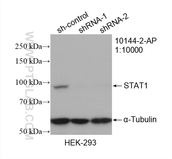
- Validé par KD/KO
Anticorps Polyclonal de lapin anti-STAT1
STAT1 Polyclonal Antibody for WB, IHC, IF/ICC, FC (Intra), IP, ELISA
Hôte / Isotype
Lapin / IgG
Réactivité testée
Humain et plus (5)
Applications
WB, IHC, IF/ICC, FC (Intra), IP, CoIP, ChIP, RIP, ELISA
Conjugaison
Non conjugué
N° de cat : 10144-2-AP
Synonymes
Galerie de données de validation
Applications testées
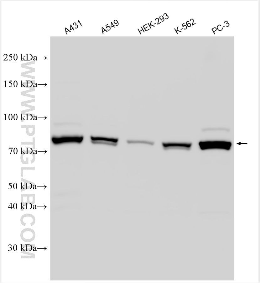
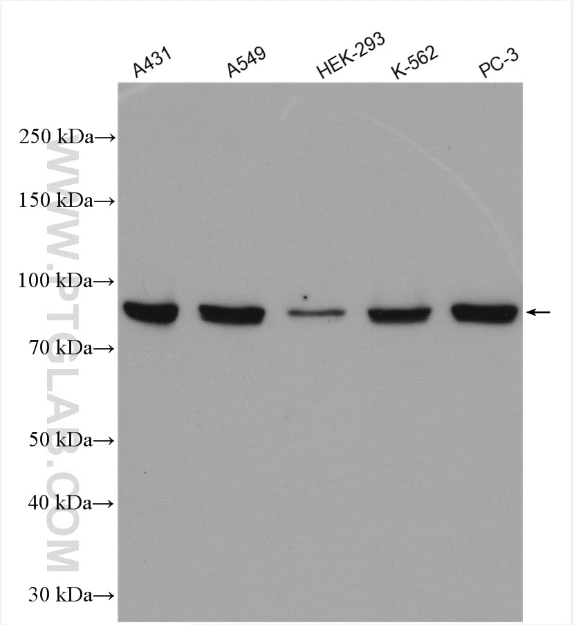
| Résultats positifs en WB | cellules A431, cellules A549, cellules HEK-293, cellules K-562, cellules PC-3 |
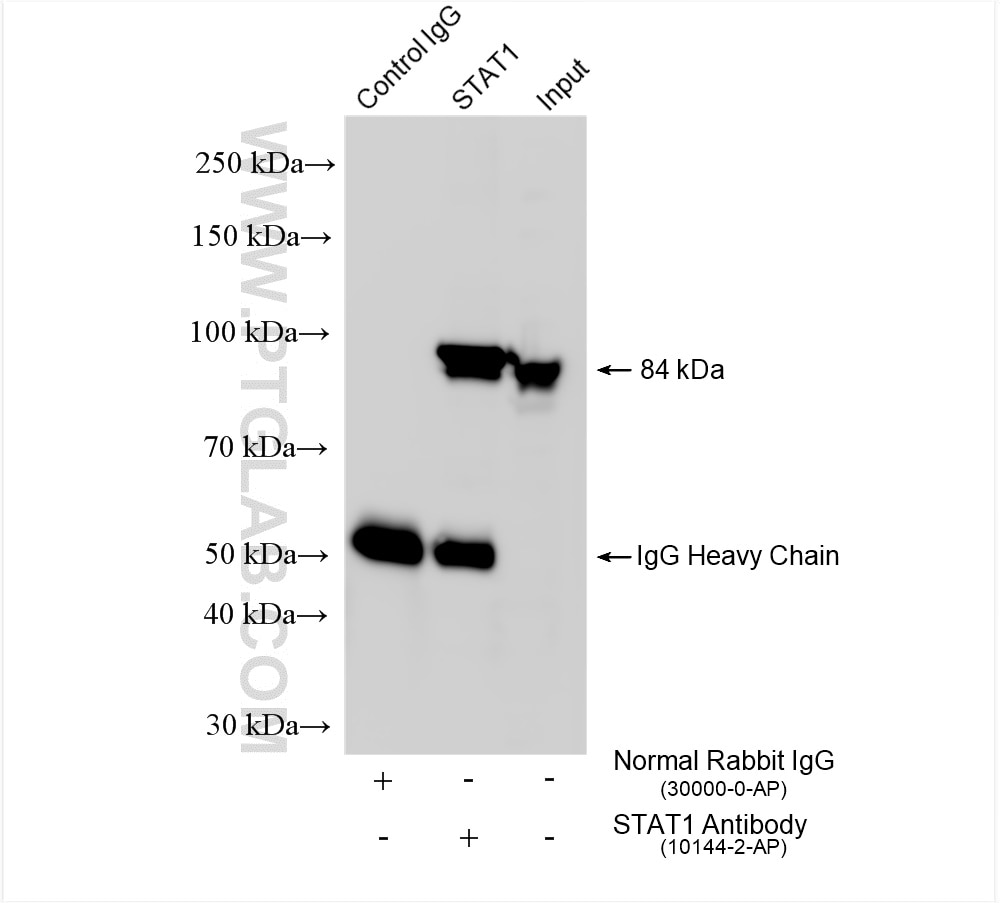
| Résultats positifs en IP | cellules PC-3, |
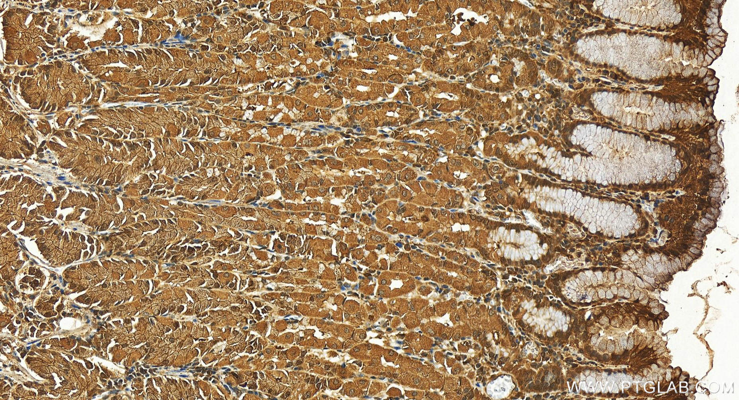
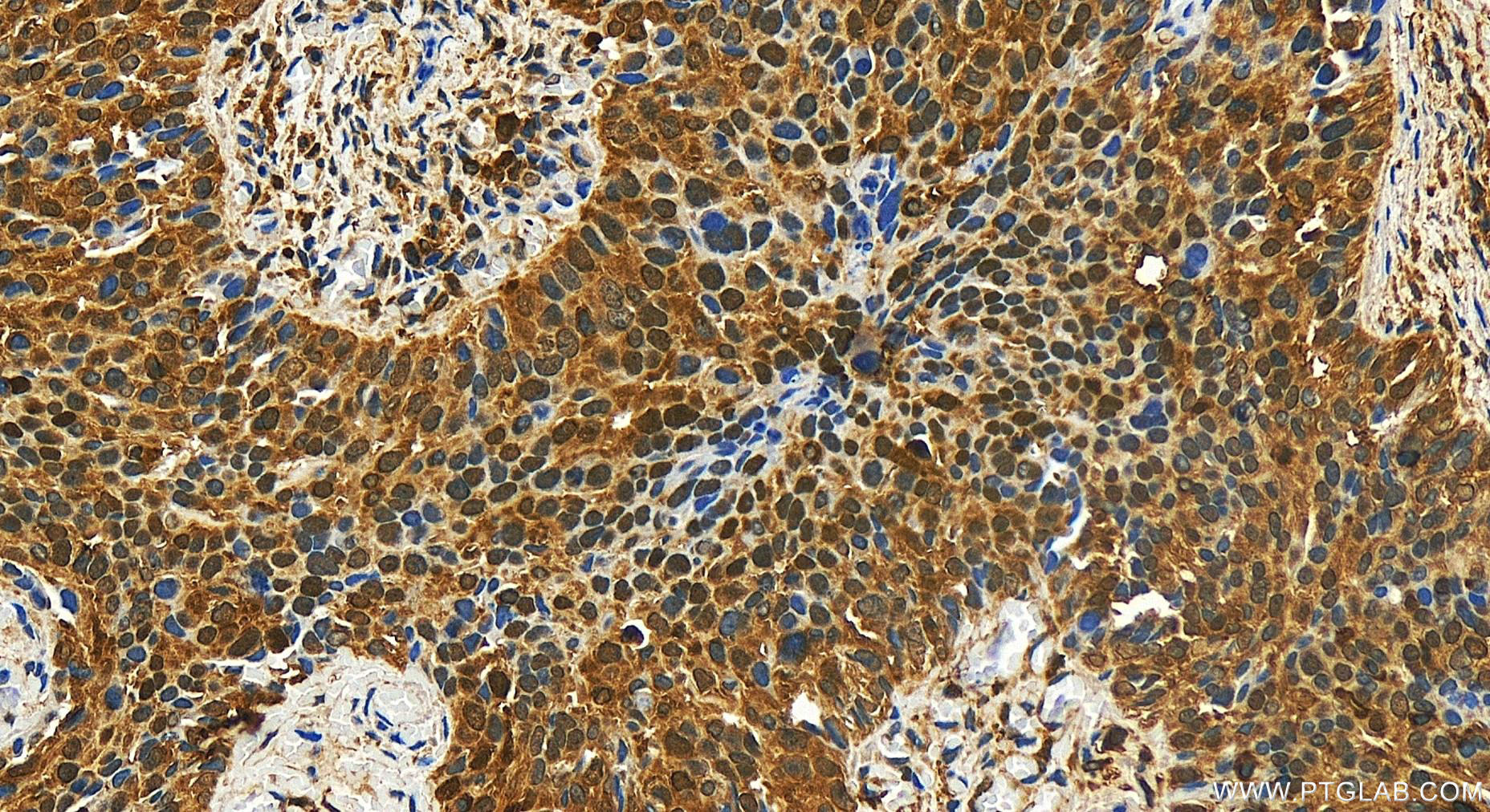
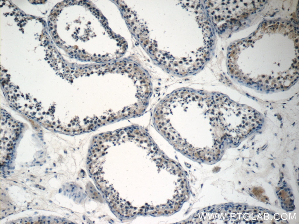
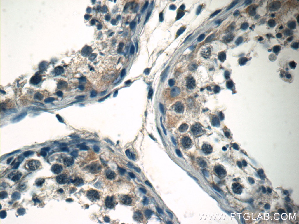
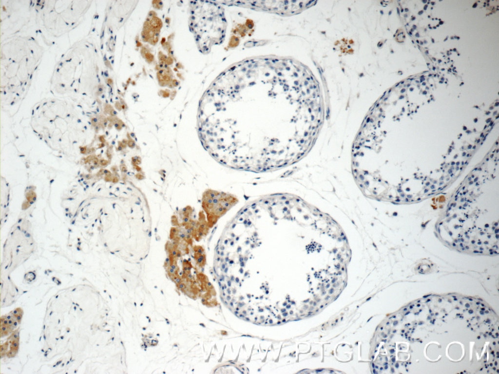
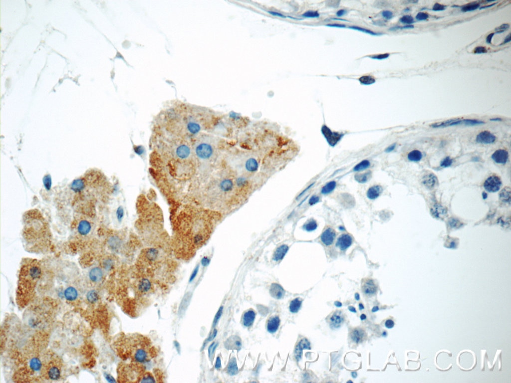
| Résultats positifs en IHC | tissu d'estomac humain, tissu testiculaire humain il est suggéré de démasquer l'antigène avec un tampon de TE buffer pH 9.0; (*) À défaut, 'le démasquage de l'antigène peut être 'effectué avec un tampon citrate pH 6,0. |
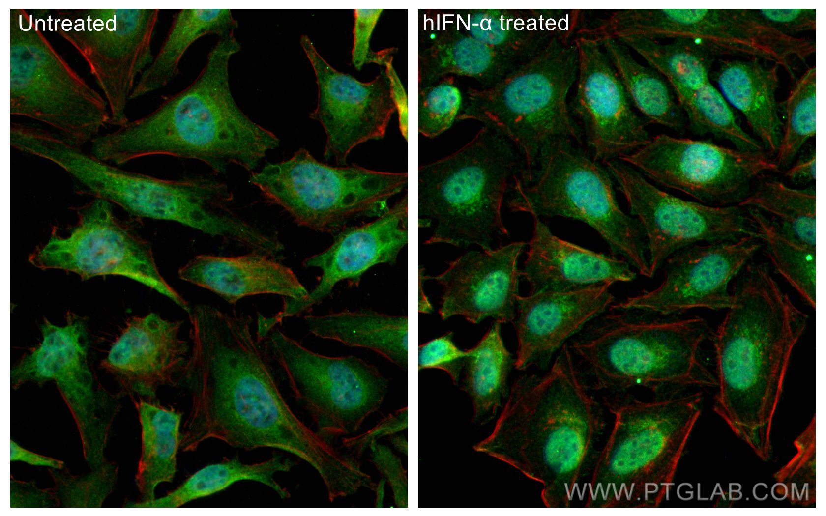
| Résultats positifs en IF/ICC | IFN alpha treated HeLa cells, |
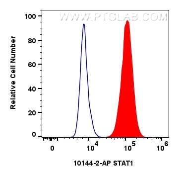
| Résultats positifs en FC (Intra) | cellules MCF-7, |
Dilution recommandée
| Application | Dilution |
|---|---|
| Western Blot (WB) | WB : 1:2000-1:10000 |
| Immunoprécipitation (IP) | IP : 0.5-4.0 ug for 1.0-3.0 mg of total protein lysate |
| Immunohistochimie (IHC) | IHC : 1:200-1:800 |
| Immunofluorescence (IF)/ICC | IF/ICC : 1:200-1:800 |
| Flow Cytometry (FC) (INTRA) | FC (INTRA) : 0.40 ug per 10^6 cells in a 100 µl suspension |
| It is recommended that this reagent should be titrated in each testing system to obtain optimal results. | |
| Sample-dependent, check data in validation data gallery | |
Informations sur le produit
10144-2-AP cible STAT1 dans les applications de WB, IHC, IF/ICC, FC (Intra), IP, CoIP, ChIP, RIP, ELISA et montre une réactivité avec des échantillons Humain
| Réactivité | Humain |
| Réactivité citée | rat, Humain, porc, poulet, singe, souris |
| Hôte / Isotype | Lapin / IgG |
| Clonalité | Polyclonal |
| Type | Anticorps |
| Immunogène | STAT1 Protéine recombinante Ag0199 |
| Nom complet | signal transducer and activator of transcription 1, 91kDa |
| Masse moléculaire calculée | 83 kDa |
| Poids moléculaire observé | 83-87 kDa |
| Numéro d’acquisition GenBank | BC002704 |
| Symbole du gène | STAT1 |
| Identification du gène (NCBI) | 6772 |
| Conjugaison | Non conjugué |
| Forme | Liquide |
| Méthode de purification | Purification par affinité contre l'antigène |
| Tampon de stockage | PBS with 0.02% sodium azide and 50% glycerol |
| Conditions de stockage | Stocker à -20°C. Stable pendant un an après l'expédition. L'aliquotage n'est pas nécessaire pour le stockage à -20oC Les 20ul contiennent 0,1% de BSA. |
Informations générales
What is the molecular weight of STAT1? Are there any isoforms of STAT1? Is STAT1 post-translationally modified?
The molecular weight of STAT1 is 91 kDa for full-length STAT1α and 84 kDa for STAT1β, which is devoid of the C-terminal activation domain (PMID: 1502203). Phosphorylation of STAT1 regulates STAT1 activity. Phosphorylation of Tyr701 by Janus kinases (JAKs) triggers STAT1 homo- and hetero-dimerization with other STAT family members and also causes nuclear translocation, while phosphorylation of Ser727 is needed for full activation (PMID: 10851062). STAT1β lacks Ser727 and has therefore been considered to be inactive or acting in a dominant negative manner to STAT1α, although it has been further shown to be capable of transcription activation in vivo (PMID: 24710278).
What is the subcellular localization of STAT1?
STAT1 is present in the cytoplasm and in the nucleus. Translocation of STAT1 from the cytoplasm to the nucleus is required for its regulation of gene expression and is dependent on phosphorylation (see above). However, low levels of unphosphorylated STAT1, often referred to as U-STAT1, can also be found in the nucleus, where U-STAT1 regulates the stability of heterochromatin by binding to chromatin in monomeric or dimeric states (PMID: 18840523).
What is the tissue expression pattern of STAT1?
STAT1 is ubiquitously expressed.
Protocole
| Product Specific Protocols | |
|---|---|
| WB protocol for STAT1 antibody 10144-2-AP | Download protocol |
| IHC protocol for STAT1 antibody 10144-2-AP | Download protocol |
| IF protocol for STAT1 antibody 10144-2-AP | Download protocol |
| IP protocol for STAT1 antibody 10144-2-AP | Download protocol |
| Standard Protocols | |
|---|---|
| Click here to view our Standard Protocols |
Publications
| Species | Application | Title |
|---|---|---|
Nat Commun ID1 expressing macrophages support cancer cell stemness and limit CD8+ T cell infiltration in colorectal cancer | ||
Nat Commun Injectable ECM-mimetic dynamic hydrogels abolish ferroptosis-induced post-discectomy herniation through delivering nucleus pulposus progenitor cell-derived exosomes | ||
Sci Transl Med TRIB3 reduces CD8+ T cell infiltration and induces immune evasion by repressing the STAT1-CXCL10 axis in colorectal cancer. | ||
Gut Microbes Synergistic activity of Enterococcus Faecium-induced ferroptosis via expansion of IFN-γ+CD8+ T cell population in advanced hepatocellular carcinoma treated with sorafenib | ||
Small Cell-Free Extracts from Human Fat Tissue with a Hyaluronan-Based Hydrogel Attenuate Inflammation in a Spinal Cord Injury Model through M2 Microglia/Microphage Polarization. |
Avis
The reviews below have been submitted by verified Proteintech customers who received an incentive for providing their feedback.
FH Paul (Verified Customer) (11-24-2022) | Clear band at the expected size range
|
FH Alejandro (Verified Customer) (08-22-2022) | Works fine for both WB, with clear bands, and IF, which shows a high specific staining
|
FH Slavica (Verified Customer) (10-04-2021) | The antibody worked very well!!!
|
