- Phare
- Validé par KD/KO
Anticorps Polyclonal de lapin anti-TDP-43 (C-terminal)
TDP-43 (C-terminal) Polyclonal Antibody for WB, IHC, IF/ICC, IF-Fro, FC (Intra), IP, ELISA
Hôte / Isotype
Lapin / IgG
Réactivité testée
Humain, rat, souris et plus (4)
Applications
WB, IHC, IF/ICC, IF-Fro, FC (Intra), IP, CoIP, chIP, RIP, ELISA
Conjugaison
Non conjugué
N° de cat : 12892-1-AP
Synonymes
Galerie de données de validation
 antibody) at dilution of 1:30000 incubated at room temperature for 1.5 hours.")
 were subjected to SDS PAGE followed by western blot with 12892-1-AP (TDP43 antibody) at dilution of 1:1000.")
 antibody) at dilution of 1:4000 incubated at room temperature for 1.5 hours.")
 at dilution of 1:1500 incubated at room temperature for 1.5 hours.")
 (IP:12892-1-AP, 4ug; Detection:12892-1-AP 1:20000) with mouse brain tissue lysate 2120 ug.")
 (IP:12892-1-AP, 4ug; Detection:12892-1-AP 1:1000) with mouse brain tissue lysate 3000ug.")
 antibody at dilution of 1:1000 (under 10x lens). Heat mediated antigen retrieval with Tris-EDTA buffer (pH 9.0).")
 antibody) at dilution of 1:2000 (under 10x lens). Heat mediated antigen retrieval with Tris-EDTA buffer (pH 9.0).")
 antibody) at dilution of 1:1000 (under 40x lens). Heat mediated antigen retrieval with Tris-EDTA buffer (pH 9.0).")
 antibody (12892-1-AP) at dilution of 1:400 and CoraLite®488-Conjugated AffiniPure Goat Anti-Rabbit IgG(H+L) (SA00013-2).")
 fixed frozen OCT-embedded mouse brain tissue using TDP-43 (C-terminal) antibody (12892-1-AP) at dilution of 1:200 and CoraLite®488-Conjugated Goat Anti-Rabbit IgG(H+L) (SA00013-2).")
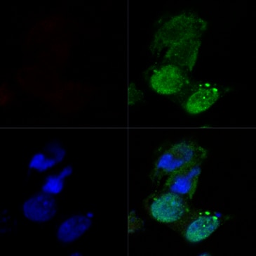
 fixed HeLa cells using TDP-43 (C-terminal) antibody (12892-1-AP) at dilution of 1:4000 and CoraLite®488-Conjugated AffiniPure Goat Anti-Rabbit IgG(H+L), CL594-Phalloidin (red).")
 fixed HeLa cells using TDP-43 (C-terminal) antibody (12892-1-AP) at dilution of 1:2400 and CoraLite®488-Conjugated AffiniPure Goat Anti-Rabbit IgG(H+L), CL594-Phalloidin (red).")
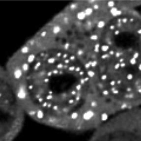
 fixed Neuro-2a cells using TDP-43 (C-terminal) antibody (12892-1-AP) at dilution of 1:8000 and CoraLite®488-Conjugated AffiniPure Goat Anti-Rabbit IgG(H+L).")
 fixed Neuro-2a cells using TDP-43 (C-terminal) antibody (12892-1-AP) at dilution of 1:8000 and CoraLite®488-Conjugated AffiniPure Goat Anti-Rabbit IgG(H+L).")
 (12892-1-AP) and CoraLite®488-Conjugated AffiniPure Goat Anti-Rabbit IgG(H+L) at dilution 1:1000 (red), or 0.4 ug Isotype Control. Cells were fixed and permeabilized with Transcription Factor Staining Buffer Kit (PF00011).")
Applications testées
| Résultats positifs en WB | cellules A549, cellules C6, cellules HeLa, cellules K-562, tissu cérébral de souris |
| Résultats positifs en IP | tissu cérébral de souris, |
| Résultats positifs en IHC | tissu cérébral de rat, tissu cérébral de souris, tissu de gliome humain il est suggéré de démasquer l'antigène avec un tampon de TE buffer pH 9.0; (*) À défaut, 'le démasquage de l'antigène peut être 'effectué avec un tampon citrate pH 6,0. |
| Résultats positifs en IF-Fro | tissu cérébral de souris, |
| Résultats positifs en IF/ICC | cellules HeLa, cellules Neuro-2a |
| Résultats positifs en FC (Intra) | cellules HeLa, |
Dilution recommandée
| Application | Dilution |
|---|---|
| Western Blot (WB) | WB : 1:5000-1:50000 |
| Immunoprécipitation (IP) | IP : 0.5-4.0 ug for 1.0-3.0 mg of total protein lysate |
| Immunohistochimie (IHC) | IHC : 1:1000-1:4000 |
| Immunofluorescence (IF)-FRO | IF-FRO : 1:50-1:500 |
| Immunofluorescence (IF)/ICC | IF/ICC : 1:2000-1:8000 |
| Flow Cytometry (FC) (INTRA) | FC (INTRA) : 0.40 ug per 10^6 cells in a 100 µl suspension |
| It is recommended that this reagent should be titrated in each testing system to obtain optimal results. | |
| Sample-dependent, check data in validation data gallery | |
Informations sur le produit
12892-1-AP cible TDP-43 (C-terminal) dans les applications de WB, IHC, IF/ICC, IF-Fro, FC (Intra), IP, CoIP, chIP, RIP, ELISA et montre une réactivité avec des échantillons Humain, rat, souris
| Réactivité | Humain, rat, souris |
| Réactivité citée | rat, Drosophile, Humain, poisson-zèbre, poulet, singe, souris |
| Hôte / Isotype | Lapin / IgG |
| Clonalité | Polyclonal |
| Type | Anticorps |
| Immunogène | Protéine recombinante |
| Nom complet | TAR DNA binding protein |
| Masse moléculaire calculée | 43 kDa |
| Poids moléculaire observé | 43-45 kDa, 35 kDa |
| Numéro d’acquisition GenBank | BC001487 |
| Symbole du gène | TDP-43 |
| Identification du gène (NCBI) | 23435 |
| Conjugaison | Non conjugué |
| Forme | Liquide |
| Méthode de purification | Purification par affinité contre l'antigène |
| Tampon de stockage | PBS with 0.02% sodium azide and 50% glycerol |
| Conditions de stockage | Stocker à -20°C. Stable pendant un an après l'expédition. L'aliquotage n'est pas nécessaire pour le stockage à -20oC Les 20ul contiennent 0,1% de BSA. |
Informations générales
Transactivation response (TAR), DNA-binding protein of 43 kDa (also known as TARDBP or TDP-43), was first isolated as a transcriptional inactivator binding to the TAR DNA element of the HIV-1 virus. Neumann et al. (2006) found that a hyperphosphorylated, ubiquitinated, and cleaved form of TARDBP, known as pathologic TDP-43, is the major component of the tau-negative and ubiquitin-positive inclusions that characterize amyotrophic lateral sclerosis (ALS) and the most common pathological subtype of frontotemporal lobar degeneration (FTLD-U). 12892-1-AP is a rabbit polyclonal antibody raised against the C-terminal amino acids of human TDP-43. This antibody recognizes the cleavage product of 20-30 kDa in addition to the native and phosphorylated forms of TDP-43. Immunohistochemical analyses of TDP-43 using this antibody detect both normal diffuse nuclear staining and insoluble inclusions in pathologic tissues. Various forms of TDP-43 exist, including 18-35 kDa of cleaved C-terminal fragments, 45-50 kDa phosphoprotein, 55 kDa glycosylated form, 75 kDa hyperphosphorylated form, and 90-300 kDa cross-linked form. (17023659,19823856,21666678,22193176)
Recently TDP-43 has been reported to be overexpressed in triple negative breast cancer (TNBC) and it may be a potential target for TNBC diagnosis and drug design. (29581274)
Protocole
| Product Specific Protocols | |
|---|---|
| WB protocol for TDP-43 (C-terminal) antibody 12892-1-AP | Download protocol |
| IHC protocol for TDP-43 (C-terminal) antibody 12892-1-AP | Download protocol |
| IF protocol for TDP-43 (C-terminal) antibody 12892-1-AP | Download protocol |
| IP protocol for TDP-43 (C-terminal) antibody 12892-1-AP | Download protocol |
| Standard Protocols | |
|---|---|
| Click here to view our Standard Protocols |
Publications
| Species | Application | Title |
|---|---|---|
Science HSP70 chaperones RNA-free TDP-43 into anisotropic intranuclear liquid spherical shells.
| ||
Nature Therapeutic reduction of ataxin-2 extends lifespan and reduces pathology in TDP-43 mice. | ||
Nat Med The inhibition of TDP-43 mitochondrial localization blocks its neuronal toxicity. |
Avis
The reviews below have been submitted by verified Proteintech customers who received an incentive for providing their feedback.
FH Emilie (Verified Customer) (09-24-2025) | I used it both in WB (1:1000) and IF (1:200), and it performed as expected.
|
FH Manon (Verified Customer) (09-23-2025) | The antibody produced clear, specific staining by immunofluorescence and also detected the protein at the expected size by western blot.
|
FH Xiaochen (Verified Customer) (11-11-2024) | sensitive and specific
 |
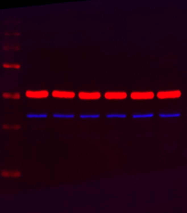
FH Scott (Verified Customer) (10-22-2024) | 10µg of protein was loaded and antibody was incubated overnight at 4oC following a total protein stain. The band appeared at the expected size, blue bar, with Alpha-tubulin internal control (red band - 66031-1-Ig). Precision plus protein standard ladder #1610373.
 |
FH Parijat (Verified Customer) (09-09-2023) | Good antibody
 |
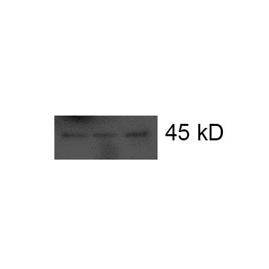
FH Xin (Verified Customer) (01-23-2022) | Good performance in WB (around 45 kD)
 |
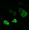
FH Azita (Verified Customer) (06-17-2021) | Immunocytochemistry labelling of (4% PFA) fixed NSC-34 cells by TDP-43 (C-terminal) Polyclonal antibody at dilution of 1:500 showed strong labelling.
|
FH Jacob (Verified Customer) (03-25-2021) | Good for IF staining. It worked for cells fixation with 4% PFA.
 |
FH David (Verified Customer) (01-13-2020) | Good for both immunofluorescence and immunoblotting. Single band in control cells in the latter, but can detect stress induced C-terminal fragments.
|
FH Alinda (Verified Customer) (02-28-2019) | Good antibody
|
FH Noemi (Verified Customer) (01-24-2019) | really good antibody, it detects also the cleavage product of TDP43 in WB.
|

