- Phare
- Validé par KD/KO
Anticorps Polyclonal de lapin anti-TLR4
TLR4 Polyclonal Antibody for WB, IHC, ELISA
Hôte / Isotype
Lapin / IgG
Réactivité testée
Humain, rat, souris et plus (7)
Applications
WB, IHC, IF, IP, CoIP, ChIP, ELISA, Microtiter plate binding assay
Conjugaison
Non conjugué
N° de cat : 19811-1-AP
Synonymes
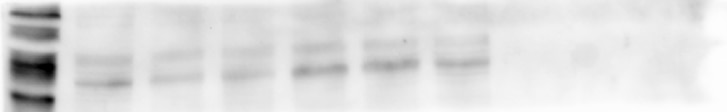
Galerie de données de validation
 at dilution of 1:2000 incubated at room temperature for 1.5 hours.")
 at dilution of 1:1000 incubated at room temperature for 1.5 hours.")
 at dilution of 1:200 (under 10x lens).")
 at dilution of 1:200 (under 40x lens).")
 at dilution of 1:200 (under 10x lens).")
 at dilution of 1:200 (under 40x lens).")
 at dilution of 1:200 (under 10x lens. Heat mediated antigen retrieval with Tris-EDTA buffer (pH 9.0).")
 at dilution of 1:200 (under 40x lens. Heat mediated antigen retrieval with Tris-EDTA buffer (pH 9.0).")
 at dilution of 1:200 (under 10x lens). Heat mediated antigen retrieval with Tris-EDTA buffer (pH 9.0).")
 at dilution of 1:200 (under 40x lens). Heat mediated antigen retrieval with Tris-EDTA buffer (pH 9.0).")
Applications testées
| Résultats positifs en WB | tissu hépatique de souris, cellules NIH/3T3, cellules RAW 264.7 |
| Résultats positifs en IHC | tissu placentaire humain, tissu cérébral de souris, tissu de cancer du foie humain, tissu de côlon humain il est suggéré de démasquer l'antigène avec un tampon de TE buffer pH 9.0; (*) À défaut, 'le démasquage de l'antigène peut être 'effectué avec un tampon citrate pH 6,0. |
Dilution recommandée
| Application | Dilution |
|---|---|
| Western Blot (WB) | WB : 1:1000-1:4000 |
| Immunohistochimie (IHC) | IHC : 1:100-1:400 |
| It is recommended that this reagent should be titrated in each testing system to obtain optimal results. | |
| Sample-dependent, check data in validation data gallery | |
Informations sur le produit
19811-1-AP cible TLR4 dans les applications de WB, IHC, IF, IP, CoIP, ChIP, ELISA, Microtiter plate binding assay et montre une réactivité avec des échantillons Humain, rat, souris
| Réactivité | Humain, rat, souris |
| Réactivité citée | rat, bovin, canin, Humain, Lapin, porc, poulet, souris, Hamster, duck |
| Hôte / Isotype | Lapin / IgG |
| Clonalité | Polyclonal |
| Type | Anticorps |
| Immunogène | TLR4 Protéine recombinante Ag13841 |
| Nom complet | toll-like receptor 4 |
| Masse moléculaire calculée | 839 aa, 96 kDa |
| Poids moléculaire observé | 90-110 kDa |
| Numéro d’acquisition GenBank | BC117422 |
| Symbole du gène | TLR4 |
| Identification du gène (NCBI) | 7099 |
| Conjugaison | Non conjugué |
| Forme | Liquide |
| Méthode de purification | Purification par affinité contre l'antigène |
| Tampon de stockage | PBS with 0.02% sodium azide and 50% glycerol |
| Conditions de stockage | Stocker à -20°C. Stable pendant un an après l'expédition. L'aliquotage n'est pas nécessaire pour le stockage à -20oC Les 20ul contiennent 0,1% de BSA. |
Informations générales
TLR4, also named CD284, belongs to the Toll-like receptor family. TLR4 interacts with LY96 and CD14 to mediate the innate immune response to bacterial lipopolysaccharide (LPS). TLR4 acts via MYD88, TIRAP, and TRAF6, leading to NF-kB activation, cytokine secretion, and the inflammatory response. Three alternatively spliced transcript variants that encode different protein isoforms have been described. The calculated molecular weights of the three TLR4 isoforms are 96, 91, and 73 kDa.
Protocole
| Product Specific Protocols | |
|---|---|
| WB protocol for TLR4 antibody 19811-1-AP | Download protocol |
| IHC protocol for TLR4 antibody 19811-1-AP | Download protocol |
| Standard Protocols | |
|---|---|
| Click here to view our Standard Protocols |
Publications
| Species | Application | Title |
|---|---|---|
Adv Mater Biomimetic Immunosuppressive Exosomes that Inhibit Cytokine Storms Contribute to the Alleviation of Sepsis. | ||
Adv Sci (Weinh) Turmeric-Derived Nanoparticles Functionalized Aerogel Regulates Multicellular Networks to Promote Diabetic Wound Healing | ||
J Clin Invest Longistatin in tick saliva blocks advanced glycation end-product receptor activation. | ||
Gut Microbes Fusobacterium nucleatum promotes esophageal squamous cell carcinoma progression and chemoresistance by enhancing the secretion of chemotherapy-induced senescence-associated secretory phenotype via activation of DNA damage response pathway | ||
Nat Commun TLR4 signalling via Piezo1 engages and enhances the macrophage mediated host response during bacterial infection. | ||
Nat Commun Alarmin-painted exosomes elicit persistent antitumor immunity in large established tumors in mice.
|
Avis
The reviews below have been submitted by verified Proteintech customers who received an incentive for providing their feedback.
FH Sara (Verified Customer) (10-25-2020) | B cells (mouse) were lysated in RIPA buffer with protein and phosphatase inhibitors. 10 µg of protein per lane were loaded in a 10% PAGE-SDS gel. Proteins were transferred to a PVFD membrane (Turbo Blot system) and incubated overnight with 1:1000 of anti-TLR4, followed by anti-rabbit-HRP (1:10.000) on the next day. Two bands of around 70 kDa are detected.
 |

