Anticorps Polyclonal de lapin anti-TMEM119
TMEM119 Polyclonal Antibody for IHC, IF-P, FC (Intra), ELISA
Hôte / Isotype
Lapin / IgG
Réactivité testée
Humain, souris
Applications
WB, IHC, IF-P, FC (Intra), ELISA
Conjugaison
Non conjugué
N° de cat : 27585-1-AP
Synonymes
Galerie de données de validation
 at dilution of 1:800 (under 40x lens). Heat mediated antigen retrieval with Tris-EDTA buffer (pH 9.0).")
 fixed paraffin-embedded mouse brain tissue using TMEM119 antibody (27585-1-AP) at dilution of 1:400 and CoraLite®488-Conjugated Goat Anti-Rabbit IgG(H+L) (SA00013-2). Heat mediated antigen retrieval with Tris-EDTA buffer (pH 9.0).")
 fixed paraffin-embedded mouse liver tissue using TMEM119 antibody (27585-1-AP) at dilution of 1:200 and Multi-rAb CoraLite ® Plus 488-Goat Anti-Rabbit Recombinant Secondary Antibody (H+L) (RGAR002). Heat mediated antigen retrieval with Tris-EDTA buffer (pH 9.0).")
 and CoraLite®488-Conjugated AffiniPure Goat Anti-Rabbit IgG(H+L) (SA00013-2)(red), or 0.4 ug Isotype Control (blue). Cells were fixed with 4% PFA and permeabilized with Flow Cytometry Perm Buffer (PF00011-C).")
Applications testées
| Résultats positifs en IHC | tissu cérébral de souris, il est suggéré de démasquer l'antigène avec un tampon de TE buffer pH 9.0; (*) À défaut, 'le démasquage de l'antigène peut être 'effectué avec un tampon citrate pH 6,0. |
| Résultats positifs en IF-P | tissu cérébral de souris, tissu hépatique de souris |
| Résultats positifs en FC (Intra) | cellules HEK-293, |
Dilution recommandée
| Application | Dilution |
|---|---|
| Immunohistochimie (IHC) | IHC : 1:400-1:1600 |
| Immunofluorescence (IF)-P | IF-P : 1:200-1:800 |
| Flow Cytometry (FC) (INTRA) | FC (INTRA) : 0.40 ug per 10^6 cells in a 100 µl suspension |
| It is recommended that this reagent should be titrated in each testing system to obtain optimal results. | |
| Sample-dependent, check data in validation data gallery | |
Applications publiées
| WB | See 5 publications below |
| IHC | See 2 publications below |
| IF | See 13 publications below |
| FC | See 1 publications below |
Informations sur le produit
27585-1-AP cible TMEM119 dans les applications de WB, IHC, IF-P, FC (Intra), ELISA et montre une réactivité avec des échantillons Humain, souris
| Réactivité | Humain, souris |
| Réactivité citée | Humain, souris |
| Hôte / Isotype | Lapin / IgG |
| Clonalité | Polyclonal |
| Type | Anticorps |
| Immunogène | TMEM119 Protéine recombinante Ag26269 |
| Nom complet | transmembrane protein 119 |
| Masse moléculaire calculée | 29 kDa |
| Poids moléculaire observé | 45 kDa |
| Numéro d’acquisition GenBank | NM_181724 |
| Symbole du gène | TMEM119 |
| Identification du gène (NCBI) | 338773 |
| Conjugaison | Non conjugué |
| Forme | Liquide |
| Méthode de purification | Purification par affinité contre l'antigène |
| Tampon de stockage | PBS with 0.02% sodium azide and 50% glycerol |
| Conditions de stockage | Stocker à -20°C. Stable pendant un an après l'expédition. L'aliquotage n'est pas nécessaire pour le stockage à -20oC Les 20ul contiennent 0,1% de BSA. |
Informations générales
TMEM119 immunohistochemistry might provide a useful tool for investigating the biology and pathology of human microglia(PMID: 26250788). Microglia can be detected clearly using Catalog#27585-1-AP.
Protocole
| Product Specific Protocols | |
|---|---|
| IHC protocol for TMEM119 antibody 27585-1-AP | Download protocol |
| IF protocol for TMEM119 antibody 27585-1-AP | Download protocol |
| FC protocol for TMEM119 antibody 27585-1-AP | Download protocol |
| Standard Protocols | |
|---|---|
| Click here to view our Standard Protocols |
Publications
| Species | Application | Title |
|---|---|---|
Sci Adv Single-cell analysis of human basal cell carcinoma reveals novel regulators of tumor growth and the tumor microenvironment. | ||
Nat Commun P-selectin axis plays a key role in microglia immunophenotype and glioblastoma progression. | ||
Glia Transmembrane protein 119 is neither a specific nor a reliable marker for microglia. | ||
Pharmacol Res MSCs-extracellular vesicles attenuated neuroinflammation, synapse damage and microglial phagocytosis after hypoxia-ischemia injury by preventing osteopontin expression. | ||
Front Immunol Didymin Suppresses Microglia Pyroptosis and Neuroinflammation Through the Asc/Caspase-1/GSDMD Pathway Following Experimental Intracerebral Hemorrhage. | ||
Neurochem Res BMP7 Attenuates Neuroinflammation after Spinal Cord Injury by Suppressing the Microglia Activation and Inducing Microglial Polarization Via the STAT3 Pathway |
Avis
The reviews below have been submitted by verified Proteintech customers who received an incentive for providing their feedback.
FH Marion (Verified Customer) (07-15-2025) | Immunohistofluorescence (PFA-fixed cryosections) image of TMEM119 stained murine brain. Blocking step : 10 % Donkey Serum + 1% BSA + 0.02% Triton as blocking agent for 2 hours at RT Primary antiboby incubation time : 1 night / 4°C Secondary antibody : AlexaFluor 568 (ab175470 ; Abcam) ; 1/200 This antibody does not seem to work under these conditions.
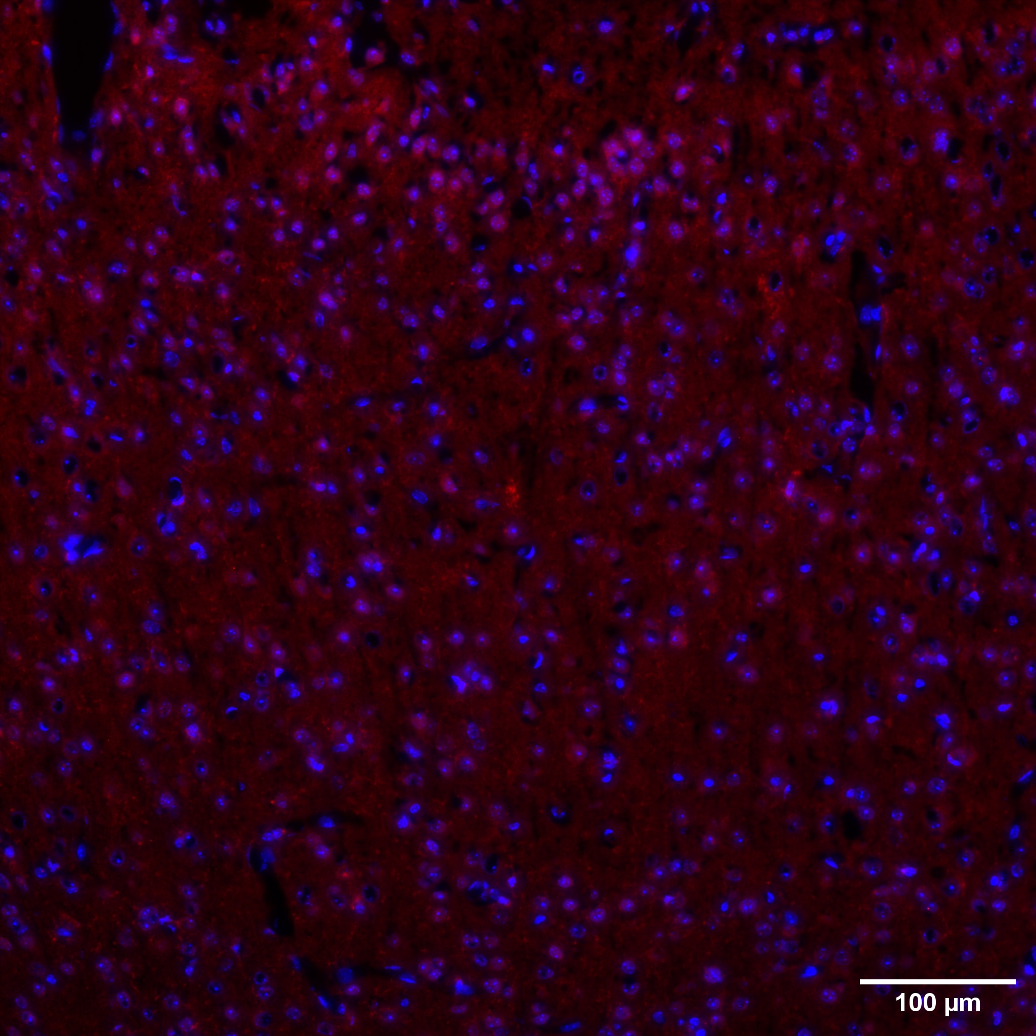 |
FH Hannes (Verified Customer) (07-27-2020) | Immunohistochemistry (Formalin/PFA-fixed paraffin-embedded sections)IHC-P image of anti-TMEM119 (27585-1-AP) stained murine and human brain. The tissue was paraffin-embedded, formalin fixed. The tissue was then incubated with EnVision+ Single Reagents (HRP. Rabbit), undiluted for 1 hour at room temperature.
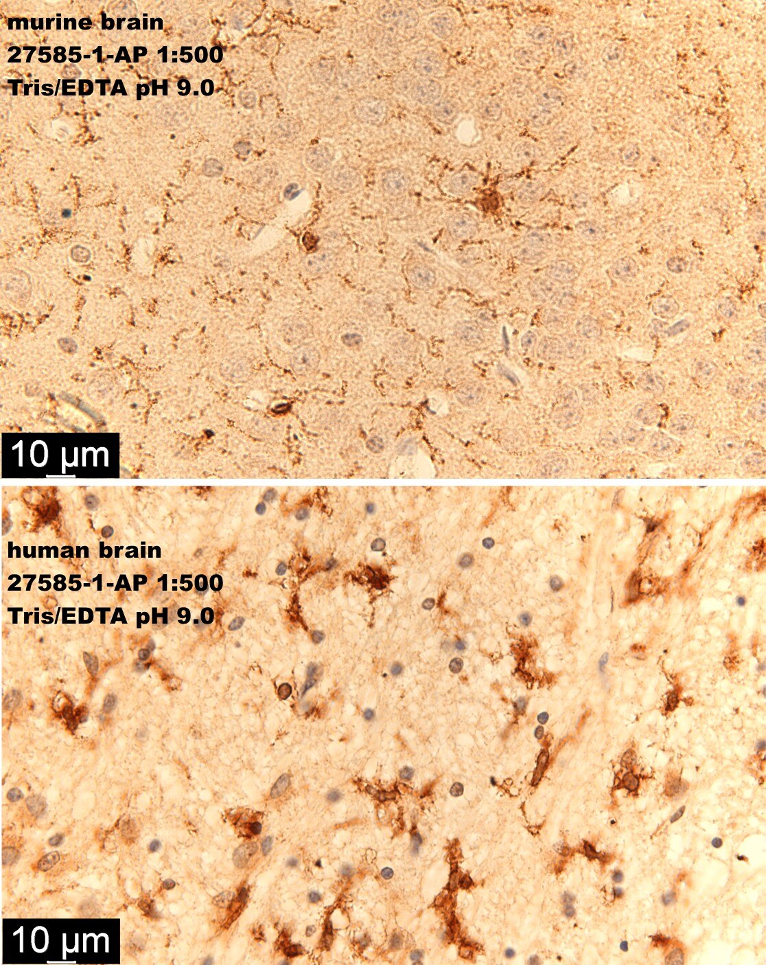 |

