Anticorps Polyclonal de lapin anti-Beta Tubulin
Beta Tubulin Polyclonal Antibody for WB, IHC, IF/ICC, IF-P, FC (Intra), IP, ELISA
Hôte / Isotype
Lapin / IgG
Réactivité testée
Humain, rat, souris et plus (6)
Applications
WB, IHC, IF/ICC, IF-P, FC (Intra), IP, CoIP, ELISA
Conjugaison
Non conjugué
N° de cat : 10094-1-AP
Synonymes
 at dilution of 1:6000 incubated at room temperature for 1.5 hours.")
 at dilution of 1:2500 incubated at room temperature for 1.5 hours.")
 at dilution of 1:1000 incubated at room temperature for 1.5 hours.")
 with mouse brain tissue lysate 7500ug.")
 at dilution of 1:200 (under 10x lens). Heat mediated antigen retrieval with Tris-EDTA buffer (pH 9.0).")
 at dilution of 1:200 (under 40x lens). Heat mediated antigen retrieval with Tris-EDTA buffer (pH 9.0).")
 at dilution of 1:50 (under 10x lens).")
 at dilution of 1:50 (under 40x lens).")
 at dilution of 1:50 (under 40x lens).")
 at dilution of 1:50 (under 10x lens).")
 at dilution of 1:200 (under 10x lens. Heat mediated antigen retrieval with Tris-EDTA buffer (pH 9.0).")
 at dilution of 1:200 (under 40x lens. Heat mediated antigen retrieval with Tris-EDTA buffer (pH 9.0).")
 at dilution of 1:200 (under 10x lens).")
 at dilution of 1:200 (under 40x lens).")
 fixed paraffin-embedded mouse eye tissue using Beta Tubulin antibody (10094-1-AP) at dilution of 1:200 and CoraLite®488-Conjugated Goat Anti-Rabbit IgG(H+L) (SA00013-2). Heat mediated antigen retrieval with Tris-EDTA buffer (pH 9.0).")
 fixed HepG2 cells using 10094-1-AP (beta Tubulin antibody) at dilution of 1:50 and Alexa Fluor 488-conjugated AffiniPure Goat Anti-Rabbit IgG(H+L).")
 fixed HeLa cells using Beta Tubulin antibody (10094-1-AP) at dilution of 1:400 and CoraLite®488-Conjugated AffiniPure Goat Anti-Rabbit IgG(H+L) (SA00013-2).")
 and CoraLite®488-Conjugated Goat Anti-Rabbit IgG(H+L) (SA00013-2)(red), or 0.25 ug rabbit IgG isotype control (blue). Cells were fixed with 4% PFA and permeabilized with Flow Cytometry Perm Buffer (PF00011-C).")
"Beta Tubulin Antibodies" Comparison
View side-by-side comparison of Beta Tubulin antibodies from other vendors to find the one that best suits your research needs.
Applications testées
| Résultats positifs en WB | cellules U-251, cellules HEK-293, tissu cérébral de rat, tissu cérébral de souris, tissu rénal de rat, tissu rénal de souris |
| Résultats positifs en IP | tissu cérébral de souris |
| Résultats positifs en IHC | tissu de côlon humain, tissu cérébral de souris, tissu de cervelet humain, tissu testiculaire de rat il est suggéré de démasquer l'antigène avec un tampon de TE buffer pH 9.0; (*) À défaut, 'le démasquage de l'antigène peut être 'effectué avec un tampon citrate pH 6,0. |
| Résultats positifs en IF-P | tissu oculaire de souris, |
| Résultats positifs en IF/ICC | cellules HeLa, cellules HepG2 |
| Résultats positifs en FC (Intra) | cellules HepG2, |
Dilution recommandée
| Application | Dilution |
|---|---|
| Western Blot (WB) | WB : 1:2000-1:12000 |
| Immunoprécipitation (IP) | IP : 0.5-4.0 ug for 1.0-3.0 mg of total protein lysate |
| Immunohistochimie (IHC) | IHC : 1:50-1:500 |
| Immunofluorescence (IF)-P | IF-P : 1:50-1:500 |
| Immunofluorescence (IF)/ICC | IF/ICC : 1:200-1:800 |
| Flow Cytometry (FC) (INTRA) | FC (INTRA) : 0.25 ug per 10^6 cells in a 100 µl suspension |
| It is recommended that this reagent should be titrated in each testing system to obtain optimal results. | |
| Sample-dependent, check data in validation data gallery | |
Applications publiées
| WB | See 1024 publications below |
| IHC | See 2 publications below |
| IF | See 34 publications below |
| IP | See 2 publications below |
| CoIP | See 3 publications below |
Informations sur le produit
10094-1-AP cible Beta Tubulin dans les applications de WB, IHC, IF/ICC, IF-P, FC (Intra), IP, CoIP, ELISA et montre une réactivité avec des échantillons Humain, rat, souris
| Réactivité | Humain, rat, souris |
| Réactivité citée | rat, Chèvre, Humain, Lapin, poisson-zèbre, poulet, souris, Hamster, fish |
| Hôte / Isotype | Lapin / IgG |
| Clonalité | Polyclonal |
| Type | Anticorps |
| Immunogène | Beta Tubulin Protéine recombinante Ag0136 |
| Nom complet | tubulin, beta 3 |
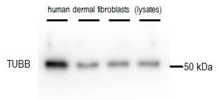
| Masse moléculaire calculée | 50 kDa |
| Poids moléculaire observé | 50-55 kDa |
| Numéro d’acquisition GenBank | BC000748 |
| Symbole du gène | TUBB3 |
| Identification du gène (NCBI) | 10381 |
| Conjugaison | Non conjugué |
| Forme | Liquide |
| Méthode de purification | Purification par affinité contre l'antigène |
| Tampon de stockage | PBS with 0.02% sodium azide and 50% glycerol |
| Conditions de stockage | Stocker à -20°C. Stable pendant un an après l'expédition. L'aliquotage n'est pas nécessaire pour le stockage à -20oC Les 20ul contiennent 0,1% de BSA. |
Informations générales
There are five tubulins in human cells: alpha, beta, gamma, delta, and epsilon. Tubulins are conserved across species. They form heterodimers, which multimerize to form a microtubule filament. An alpha and beta tubulin heterodimer is the basic structural unit of microtubules. The heterodimer does not come apart, once formed. The alpha and beta tubulins, which are each about 55 kDa MW, are homologous but not identical. Alpha, beta, and gamma tubulins have all been used as loading controls. Tubulin expression may vary according to resistance to antimicrobial and antimitotic drugs.
Protocole
| Product Specific Protocols | |
|---|---|
| WB protocol for Beta Tubulin antibody 10094-1-AP | Download protocol |
| IHC protocol for Beta Tubulin antibody 10094-1-AP | Download protocol |
| IF protocol for Beta Tubulin antibody 10094-1-AP | Download protocol |
| IP protocol for Beta Tubulin antibody 10094-1-AP | Download protocol |
| Standard Protocols | |
|---|---|
| Click here to view our Standard Protocols |
Publications
| Species | Application | Title |
|---|---|---|
Nat Biotechnol Magnify is a universal molecular anchoring strategy for expansion microscopy | ||
Nat Nanotechnol Long-term pulmonary exposure to multi-walled carbon nanotubes promotes breast cancer metastatic cascades. | ||
Cell Metab TMEM41B acts as an ER scramblase required for lipoprotein biogenesis and lipid homeostasis. | ||
Cell Metab Ejection of damaged mitochondria and their removal by macrophages ensure efficient thermogenesis in brown adipose tissue. | ||
Nat Cell Biol Single-cell multi-omics profiling of human preimplantation embryos identifies cytoskeletal defects during embryonic arrest | ||
Avis
The reviews below have been submitted by verified Proteintech customers who received an incentive for providing their feedback.
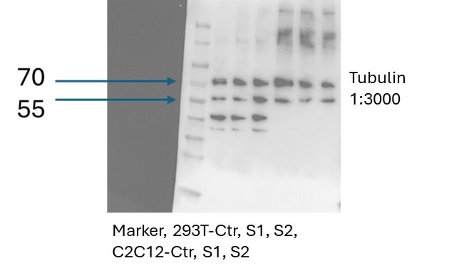
FH Dan (Verified Customer) (08-25-2025) | transfection done on 293T and C2C12 cells and 40ug loaded for western blot. two major bands seen at 56 and 72KD
 |
FH PK (Verified Customer) (08-14-2024) | excellent
 |
FH Susanne (Verified Customer) (11-25-2022) |
 |
FH Macarena (Verified Customer) (10-07-2022) | excellent results. No non specific binding. The loading control of choice in my lab
|
FH parisa (Verified Customer) (12-03-2021) | We had the anti-mouse antibody and tried this anti-rabbit antibody which worked very well and will be helpful for us.
|
FH Azita (Verified Customer) (06-02-2021) | Immunohistochemistry labelling of (4% PFA) fixed mice spinal cord tissues using Beta Tubulin Polyclonal Antibody at dilution of 1:50 showed strong labelling.
|
FH Elise (Verified Customer) (12-27-2020) | Works perfectly well!
|

