- Phare
- Validé par KD/KO
Anticorps Polyclonal de lapin anti-WT1
WT1 Polyclonal Antibody for WB, IF/ICC, FC (Intra), IP, ELISA
Hôte / Isotype
Lapin / IgG
Réactivité testée
Humain, rat, souris et plus (3)
Applications
WB, IF/ICC, FC (Intra), IP, chIP, ELISA
Conjugaison
Non conjugué
N° de cat : 12609-1-AP
Synonymes
Galerie de données de validation
Applications testées
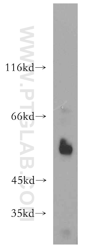
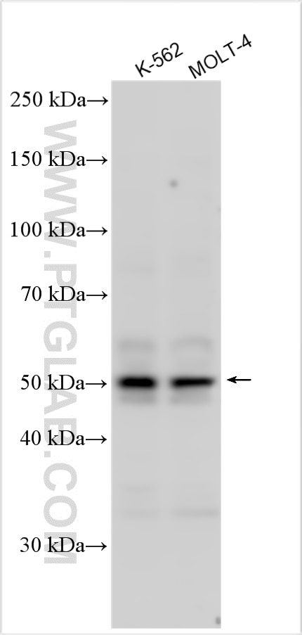
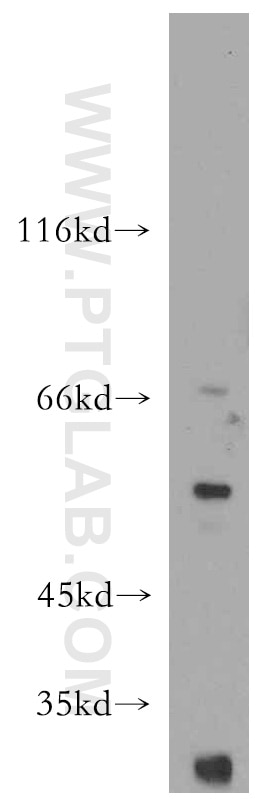
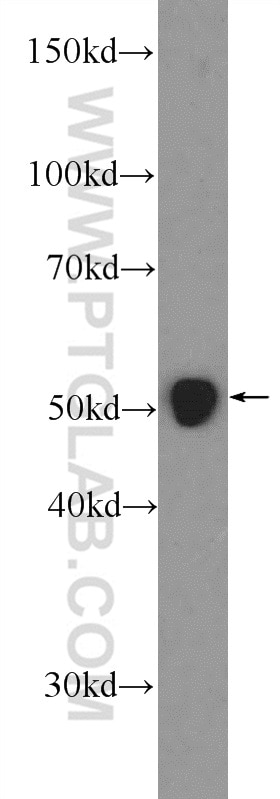
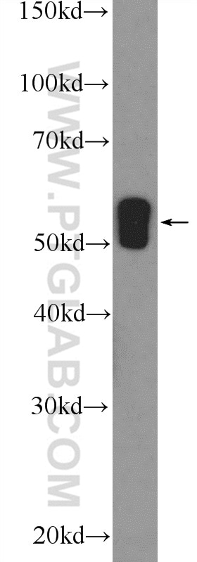
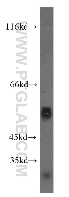
| Résultats positifs en WB | cellules K-562, cellules A431, cellules MCF-7, tissu rénal de rat, tissu rénal de souris |
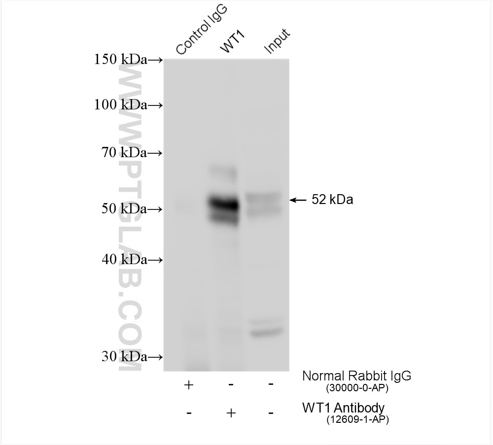
| Résultats positifs en IP | cellules K-562, |
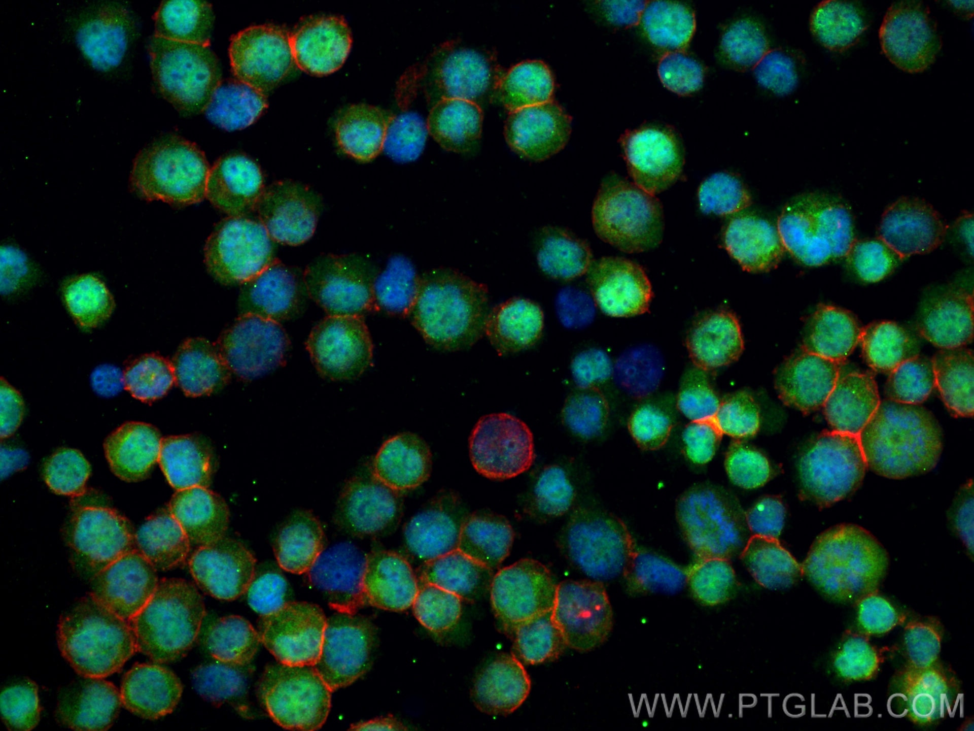
| Résultats positifs en IF/ICC | cellules K-562, |
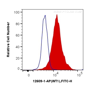
| Résultats positifs en FC (Intra) | cellules K-562, |
Dilution recommandée
| Application | Dilution |
|---|---|
| Western Blot (WB) | WB : 1:500-1:1000 |
| Immunoprécipitation (IP) | IP : 0.5-4.0 ug for 1.0-3.0 mg of total protein lysate |
| Immunofluorescence (IF)/ICC | IF/ICC : 1:50-1:500 |
| Flow Cytometry (FC) (INTRA) | FC (INTRA) : 0.40 ug per 10^6 cells in a 100 µl suspension |
| It is recommended that this reagent should be titrated in each testing system to obtain optimal results. | |
| Sample-dependent, check data in validation data gallery | |
Applications publiées
| KD/KO | See 3 publications below |
| WB | See 29 publications below |
| IHC | See 1 publications below |
| IF | See 19 publications below |
| ChIP | See 4 publications below |
Informations sur le produit
12609-1-AP cible WT1 dans les applications de WB, IF/ICC, FC (Intra), IP, chIP, ELISA et montre une réactivité avec des échantillons Humain, rat, souris
| Réactivité | Humain, rat, souris |
| Réactivité citée | rat, bovin, Chèvre, Humain, poulet, souris |
| Hôte / Isotype | Lapin / IgG |
| Clonalité | Polyclonal |
| Type | Anticorps |
| Immunogène | WT1 Protéine recombinante Ag3337 |
| Nom complet | Wilms tumor 1 |
| Masse moléculaire calculée | 449 aa, 49 kDa, 57 kDa |
| Poids moléculaire observé | 52-55 kDa |
| Numéro d’acquisition GenBank | BC032861 |
| Symbole du gène | WT1 |
| Identification du gène (NCBI) | 7490 |
| Conjugaison | Non conjugué |
| Forme | Liquide |
| Méthode de purification | Purification par affinité contre l'antigène |
| Tampon de stockage | PBS with 0.02% sodium azide and 50% glycerol |
| Conditions de stockage | Stocker à -20°C. Stable pendant un an après l'expédition. L'aliquotage n'est pas nécessaire pour le stockage à -20oC Les 20ul contiennent 0,1% de BSA. |
Informations générales
The WT1 gene encodes a zinc finger DNA-binding protein that acts as a transcriptional activator or repressor depending on the cellular or chromosomal context, and it is required for normal formation of the genitourinary system and mesothelial tissues. WT1 inhibits apoptosis through p53 and Bcl-2 and also inhibits the differentiation of leukemic cells. Function of WT1 may be isoform-specific: isoforms lacking the KTS motif may act as transcription factors. Isoforms containing the KTS motif may bind mRNA and play a role in mRNA metabolism or splicing. Isoform 1 has lower affinity for DNA, and can bind RNA. WT1 exists some isoforms with the range of molecular weight is 33-50 kDa.
Protocole
| Product Specific Protocols | |
|---|---|
| WB protocol for WT1 antibody 12609-1-AP | Download protocol |
| IF protocol for WT1 antibody 12609-1-AP | Download protocol |
| IP protocol for WT1 antibody 12609-1-AP | Download protocol |
| Standard Protocols | |
|---|---|
| Click here to view our Standard Protocols |
Publications
| Species | Application | Title |
|---|---|---|
Mol Cell The IMiD target CRBN determines HSP90 activity toward transmembrane proteins essential in multiple myeloma. | ||
Mol Psychiatry Brain-specific Wt1 deletion leads to depressive-like behaviors in mice via the recruitment of Tet2 to modulate Epo expression.
| ||
EMBO Mol Med Inhibition of Aurora Kinase B attenuates fibroblast activation and pulmonary fibrosis. | ||
Oncogene YAP induces high-grade serous carcinoma in fallopian tube secretory epithelial cells. | ||
Oncogene USP13 promotes development and metastasis of high-grade serous ovarian carcinoma in a novel mouse model. |
Avis
The reviews below have been submitted by verified Proteintech customers who received an incentive for providing their feedback.
FH Sandrine (Verified Customer) (12-17-2021) | clean signal for IP and western blot. Good relative enrichment for ChIP assays.
|
FH K (Verified Customer) (12-20-2020) | This Ab worked well for IHC (1:200) but I could't get satisfactory results for western blotting.
|
