Anticorps Polyclonal de lapin anti-YPEL5
YPEL5 Polyclonal Antibody for WB, IHC, ELISA
Hôte / Isotype
Lapin / IgG
Réactivité testée
Humain, rat, souris et plus (1)
Applications
WB, IHC, IF, ELISA
Conjugaison
Non conjugué
N° de cat : 11730-1-AP
Synonymes
Galerie de données de validation
 at dilution of 1:2000 incubated at room temperature for 1.5 hours.")
 at dilution of 1:500 incubated at room temperature for 1.5 hours.")
 at dilution of 1:500 incubated at room temperature for 1.5 hours.")
 at dilution of 1:300 incubated at room temperature for 1.5 hours.")
 at dilution of 1:300 incubated at room temperature for 1.5 hours.")
 at dilution of 1:500 incubated at room temperature for 1.5 hours.")
 at dilution of 1:200 (under 10x lens). Heat mediated antigen retrieval with Tris-EDTA buffer (pH 9.0).")
 at dilution of 1:200 (under 40x lens). Heat mediated antigen retrieval with Tris-EDTA buffer (pH 9.0).")
Applications testées
| Résultats positifs en WB | tissu cérébral de souris, tissu cérébral de rat, tissu testiculaire de souris |
| Résultats positifs en IHC | tissu testiculaire de souris, il est suggéré de démasquer l'antigène avec un tampon de TE buffer pH 9.0; (*) À défaut, 'le démasquage de l'antigène peut être 'effectué avec un tampon citrate pH 6,0. |
Dilution recommandée
| Application | Dilution |
|---|---|
| Western Blot (WB) | WB : 1:1000-1:4000 |
| Immunohistochimie (IHC) | IHC : 1:50-1:500 |
| It is recommended that this reagent should be titrated in each testing system to obtain optimal results. | |
| Sample-dependent, check data in validation data gallery | |
Applications publiées
| WB | See 5 publications below |
| IF | See 1 publications below |
Informations sur le produit
11730-1-AP cible YPEL5 dans les applications de WB, IHC, IF, ELISA et montre une réactivité avec des échantillons Humain, rat, souris
| Réactivité | Humain, rat, souris |
| Réactivité citée | Humain, poisson-zèbre, souris |
| Hôte / Isotype | Lapin / IgG |
| Clonalité | Polyclonal |
| Type | Anticorps |
| Immunogène | YPEL5 Protéine recombinante Ag2328 |
| Nom complet | yippee-like 5 (Drosophila) |
| Masse moléculaire calculée | 121 aa, 14 kDa |
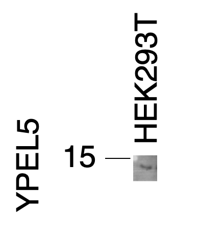
| Poids moléculaire observé | 14 kDa |
| Numéro d’acquisition GenBank | BC000836 |
| Symbole du gène | YPEL5 |
| Identification du gène (NCBI) | 51646 |
| Conjugaison | Non conjugué |
| Forme | Liquide |
| Méthode de purification | Purification par affinité contre l'antigène |
| Tampon de stockage | PBS with 0.02% sodium azide and 50% glycerol |
| Conditions de stockage | Stocker à -20°C. Stable pendant un an après l'expédition. L'aliquotage n'est pas nécessaire pour le stockage à -20oC Les 20ul contiennent 0,1% de BSA. |
Informations générales
YPEL5(Protein yippee-like 5) belongs to the yippee family and is involved in a certain cell division-related function.During cell cycle, YPEL5 protein is detected at different subcellular localizations; at interphase, it is located in the nucleus and centrosome, then it changes location sequentially to spindle poles, mitotic spindle, and spindle midzone during mitosis, and finally transferrs to midbody at cytokinesis(PMID:20580816).
Protocole
| Product Specific Protocols | |
|---|---|
| WB protocol for YPEL5 antibody 11730-1-AP | Download protocol |
| IHC protocol for YPEL5 antibody 11730-1-AP | Download protocol |
| Standard Protocols | |
|---|---|
| Click here to view our Standard Protocols |
Publications
| Species | Application | Title |
|---|---|---|
Neuron Molecular microcircuitry underlies functional specification in a basal ganglia circuit dedicated to vocal learning. | ||
Mol Oncol METTL3/YTHDF2 m6A axis accelerates colorectal carcinogenesis through epigenetically suppressing YPEL5. | ||
iScience Genome-wide CRISPR-Cas9 screen analyzed by SLIDER identifies network of repressor complexes that regulate TRIM24 | ||
bioRxiv Genome-wide screen of Mycobacterium tuberculosis- infected macrophages identified the GID/CTLH complex as a determinant of intracellular bacterial growth | ||
Nat Commun Genome-wide screen of Mycobacterium tuberculosis-infected macrophages revealed GID/CTLH complex-mediated modulation of bacterial growth |
Avis
The reviews below have been submitted by verified Proteintech customers who received an incentive for providing their feedback.
FH S (Verified Customer) (11-22-2021) | The band was there on the right MW range, but it was a bit weak. It could have been better if I used 0.2 um pore-size membrane, given that YPEL5 is a small protein.
 |

